Cushing’s Syndrome
- Chronic hypercortisolism due to excessive production of cortisol
- 20-40 years of age
- 3 times higher frequency in women
- Pituitary Cushing’s syndrome
- Adrenal Cushing’s syndrome
- Ectopic Cushing’s syndrome
- Iatrogenic Cushing’s syndrome
Pituitary Cushing’s syndrome (60-70%)
- Cushing’s disease
- Excessive ACTH, due to
- Pituitary lesion: corticotroph adenoma, multiple corticotroph microadenoma
- hypothalamic origin: excessive CRH secretion –> pituitary ACTH
- Excessive ACTH will act on adrenal cortex and cause: bilateral cortical hyperplasia
- DX: Dexamethasone
Adrenal Cushing’s syndrome (20-25%)
- Low ACTH levels, High Cortisol levels
- Disease in 1 or both adrenal glands
- Adrenal lesions: Adrenal cortical adenoma, carcinoma, cortical hyperplasia
- No response to high dose of glucocorticoids (?)
Ectopic Cushing’s syndrome (10-15%)
- High ACTH levels
- Ectopic ACTH secretions from non-endocrine tumours
- Oat cell carcinoma (lung)
- Lung cancer
- Malignant thymoma
- Pancreatic tumour
- ACTH act on adrenal cortex releasing cortisol
- Dexamethasone does not suppress cortisol secretion (acts on pituitary only)
Iatrogenic Cushing’s syndrome
- Prolonged therapeutic administration of high doses of glucocorticoids/ACTH
- Used in organ transplant recipients & autoimmune diseases.
_____________________________________________________________________
Clinical features
Refer Adrenal Steroid Hormones to understand action of glucocorticoid on body causing the clinical features.
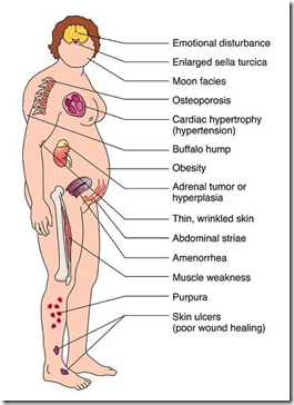
- Truncal obesity, thin extremities
- Buffalo hump (fat over shoulders)
- Moon face (rounded oedematous face)
- Muscle wasting
- Muscle weakness – hypokalemia
- Growth retardation in children & adolescents
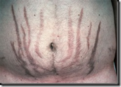
- Atrophy of subcutaneous tissue – purple striae of abdominal wall & ecchymoses. Loss of subcutaneous collagen matrix, increased skin fragility, skin peels off readily (Liddle’s sign) & subjected to fungal infection.
- Excess androgen – hirsutims, increased sebum, acne, scalp hair regression, irregular menstrual cycle
- Male: impotence, decreased libido
- Enhanced bone resorption – osteoporosis
- Polycythemia, neutrophilia, lumphopenia, eosinopenia
- hypertension, oedema & congestive heart failure
- Psychiatric: depression, psychosis, irritability
- Depressed immunity
_____________________________________________________________________
ACTH dependent (Cushing’s disease) (60%)
- pituitary origin / ectopic ACTH
- Mostly pituitary ACTH
- 15% ectopic ACTH, 50% Small cell carcionoma of the lungs
- Prominent features: hypokalemia, hypoglycemia & muscle wasting.
ACTH independent (25%)
- Adrenal origin
- Adenomas – slow growing , pure hypercortisolism (only cortisol)
- Carcinomas – larger, more cortisol precursors & adrenal androgens
- Micronodular hyperplasia – rare, common in young adults, autosomal dominant, asso. with pituitary & testicular lesions.
Diagnosis of Cushing’s syndrome
Obtain 2 late-night salivary cortisol tests (at it’s lowest level due to it’s diurnal rhythm)
Results:
- Both normal: Cushing’s unlikely
- Discordant/equivocal results – repeat or confirm with 1mg overnight low-dose dexamethasone suppression test/ UFC
- Both abnormal: Cushing’s likely – confirm with 1mg overnight low-dose dexamethasone suppression test/ UFC
Low-dose dexamethasone test explained in different post. (link)


