Evolution of Kidney Development (3 stages)
Note the image: pronephric system are at the superior (cervical region) portion, mesonephric system at the middle portion and the metanephric system at the inferior (sacral region) portion of the embryo)
Mesonephric duct opens into the urogenital sinus.
Yellow: endodermal (gut)
Blue: intermediate mesoderm (kidney & genital)
Each vertebrate’s excretory system develop:
- filtration units
- nephric duct (connected to filtration unit)
1) Pronephros (1 pair of kidneys)
- lowest verterbrates have 1 pair of kidneys
- a few nephrons (filtration unit) connected to an unbranched pronephric duct
- totally disappear at birth (human)
2) Mesonephros
- More advanced classes (fish, amphibian)
- A second pair of kidneys
- linear array of nephrons (filtration unit) connected to an unbranced mesonephric duct
- May remain at birth (part of male reproductive tract)
3)Metanephros
- Amniote classes (reptiles, birds, mammals, humans)
- Adult life human kidney
__________________________________________________________________________________________
As mentioned above, the urinary & genital system both develop from: intermediate mesoderm (along the posterior abdominal wall)
The pronephros
During 4th week, pronephros appear at the cervical region.
By the end of 4th week, pronephros disappears.
Note the intermediate column:
intermediate mesoderm where the urinary & genital system are derived from.
Note the:
- Pronephric tubule
- Primary Nephric duct
- Glomus (primitive glomerulus)
The mesonephros
At the end of 4th week, during the regression of the pronephros, the 1st tubules of mesonephros starts to appear.
By middle of 2nd month, a large mesonephros is visible.Then, the cranial tubules gradually starts degenerating.
By the end of 2nd month, most of the mesonephros disappears, but the mesonephric duct remains (only in males)
Note the intermediate mesoderm
Note the:
- Bowman’s capsule
- Mesonephric tubule
- Mesonephric duct
- Glomerulus
The pronephric components disappear.
The metanephrous
During the 5th week, the metanephros/permanent kidney appears in the sacral region of the intermediate mesoderm.
Note:
- Mesonehpric duct
- Urogenital sinus
- Ureteric bud
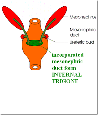
The mesonephric duct opens into the urogenital sinus.
The ureteric bud is an outgrowth from the mesonephric duct. The ureteric bud joins the metanephric blastema (also known as metanephros).
Note:
- Urogenital septum
- Cloaca
- Allantois
Portion of the hindgut (yellow) incorporated into the umbilical cord is known as the: Allantois.
Postallantoic part of the hindgut is called the: Cloaca.
The urorectal septum divides the hindgut (cloaca) into:
- Dorsal : anorectal canal (the ASS)
- Ventral: urogenital sinus (where the mesonephric duct opens into)
This is a pig embryo, note the location of the mesonephros & metanephros relative to one another.
URETERIC BUD
This image depicts the development of the metanephros to the adult kidney.
Note the ureteric bud: Dark pink
The ureteric bud elongates and become separate from the mesonephric duct (green), and is now opening into the vesico-urethral canal (pink) -> future urinary bladder.
The ureteric bud goes on penetrating the metanephric tissue (orange). The bud then dilates and forms the renal pelvis -> splits into branches (major calyces) -> each calyx forms 2 buds -> each bud forms 12/more generation of branching (2nd, 3rd, 4th orders forming the minor calyces) (1st, 5-12th orders forming the 1-3 millions of collecting tubules).
Therefore, the ureteric bud will develop the:
- Minor & major calyx
- Collecting Tubule
- Collecting duct
- Ureter
METANEPHROS
Note the:
- Metanephric tissue caps
- Renal vesicle
The metanephric mesoderm/metanephros/metanephric blastema at the end of collecting tubule are shaped in a way forming the metanephric tissue cap/metanephric mass, which will be induced by the collecting tubule to form nephrons. This metanephric tissue cap will form cell clusters, and lumen will then be formed within the cell clusters forming renal vesicle (metanephric vesicle). The renal vesicle will form a S-shaped tubule which will then develop into the bowman’s capsule. Capillaries will grow at the other end, forming glomerulus, whereby the Bowman’s capsule will fit around it. The tubules + glomerulus forms a nephron. Then the tubules of the nephron lengthen and forms the PCT, Loop of Henle & DCT.
Therefore, the metanephros will develop the:
- Bowman’s capsule
- PCT
- Loop of Henle
- DCT
SUMMARY:

*IMPORTANT FOR EXAM: The permanent kidney:
Note the regions of the functional kidney derived from the metanephric mesoderm & ureteric bud.
1) Mesonephros/metanephric blastema/metanephric mesoderm:
- Bowman’s Capsule
- Proximal Convoluted kidney
- Loop of Henle
- Distal Convoluted Kidney
2) Ureteric Bud:
- Minor & major calyx
- Collecting Tubule
- Collecting duct
- Ureter
20 nephrons open into 1 collecting tubule.
As we already know, the ureteric bud is connected with the metanephric blastema/metanephros. (which the resulting collecting duct formed from the ureteric bud will induce the metanephros to form the renal vesicles). IF the ureteric bud for some reason fails to contact/connect or induce the metanephric blastema,then the fetus will suffer from Congenital Polycystic Kidney (numerous collecting ducts surrounded by cysts/undifferentiated cells of the metanephros). There are 2 types:
1) Autosomal dominant
- Cysts from ALL parts of nephron
- More common
- Renal failure during adulthood
2) Autosomal recessive
- Multiple cysts from collecting tubule
- Progressive disorder
- Renal failure in infancy
It’s our genes that controls the formation of growth factors and determines whether the kidney develops normally or not.
It requires the epithelial-mesenchyme interaction. The signaling between these two different tissues is required to form the functional unit of the systems in which it is used.
The differentiation of the kidney involves the:
- WT1 Transcription gene (expressed by the metanephrous mesoderm) via the GDNF & HGF Growth factors
It makes the tissue component differentiate & form the nephrons (in response to the ureteric bud induction via FGF-2, BMP-7 Growth Factors).
Mutation in WT1 gene produces abnormal differention, causing Wilm’s Tumour.
_____________________________________________________________________________________________
Renal Agenesis
Renal agenesis occurs when 1 or both kidney fails to develop in fetus.
It can be caused by genetic/environmental factors:
- Untreated diabetes of the mother
The metanephric blastema/metanephros fails to mature and the ureteric bud also fails to branch/elongate. Hence they whole kidney is not formed.
There are 2 types of renal agenesis:
1) Unilateral renal agenesis
- 1 kidney missing
- remaining kidney handles the workload
- compatible with life
2) Bilateral renal agenesis
- 1:10000 live births
- both kidneys missing
- results in renal failure
It is absolutely important that the fetus develops the kidneys by the 10th week, because that will be when the fetus starts urinating and provide it’s own amniotic fluid. In normal conditions, the fetus excretes urine and form the amniotic fluid, and then engulfs it. The engulfment of the amniotic fluid creates pressure on the oesophagus and will hence develop lumen (otherwise oesophagus will remain patent/solid).
So, if there is renal agenesis, there will be less amniotic fluid formed (oligohydramnios) and hence many other organs cannot be formed normally:
- oesophageal atresia
- anorectal atresia
- hypoplastic lung
- absent sex organs
If there is polyhydramnios (too much amniotic fluid), there will be abnormal facial appearance due to the pressure on the fetus’s face.
_____________________________________________________________________________________________
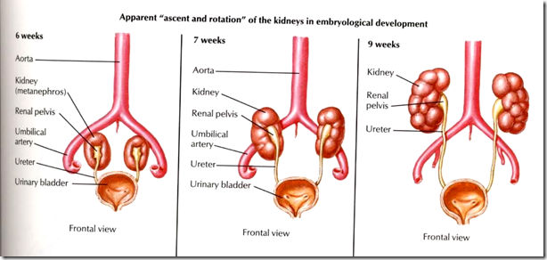
Ascent of the developed kidney
After developing in the pelvic region, it shifts to the lumbar region due to straightening of the body curvature of the fetus
In the pelvic region,
Blood supply of the kidney is from: Pelvic branch of aorta
In the lumbar region,
Blood supply of the kidney is from: Renal artery.
The lower arteries degenerates.
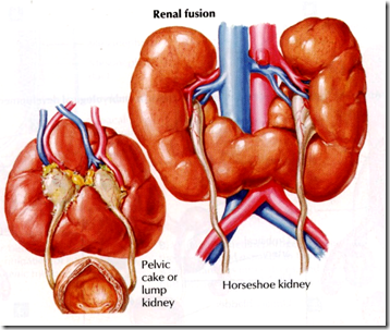
Inability of kidney to ascend:
This condition is known as pelvic kidney, where the kidney remains in the pelvic region. It can be caused by:
1) the inferior mesenteric artery blocking the ascent
2) both kidney fused at the lower lumbar level and produce HORSESHOE KIDNEY
3) If it’s fully fused at the upper and lower part, then it is known as LUMP KIDNEY
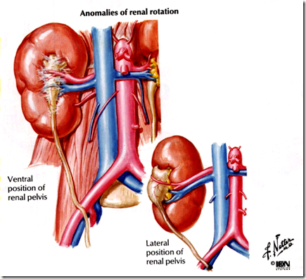
Other abnormalities with ascent of kidney:
1) Ectopic kidney
2) Abnormal duplication of the ureter (complete & incomplete)
Complete duplication of the ureter: opens into the prostate/vaginal causing hydroureter. Urine cannot be excreted/ drained therefore there will be dilatation of the ureter (hydroureter) or dilatation of the kidney (hydronephrosis).
3) Abnormal renal rotation (ventral & lateral position)
4) Supernumeric kidney (more than 2 kidneys)
SUMMARY OF DEVELOPMENTAL ANOMALIES OF THE KIDNEY
1) Formation of kidney (lack of interaction between ureteric bud & metanephros)
- Polycystic kidney
- Renal agenesis (unilateral/bilateral)
- Wilm’s Tumour
2) Ascend of kidney
- Horseshoe/Lump kidney
- Ectopic kidney
- Abnormal duplication of the ureter (com
plete/incomplete) - Abnormal rotation of the kidney
- Supranumeric kidney
_____________________________________________________________________________________________
Development of the Urinary Bladder
As discussed, during the 4th – 7th week,
The cloaca divides into the urogenital sinus & anorectal canal by urorectal septum. The tip of the urorectal septum is known as the perineal body.
With the opening of the mesonephric duct into the urogenital sinus, the urogenital sinus is divided into 3 parts:
1) Vesico-urethral canal
- forming the urinary bladder
2) Pelvic part
- forming the prostatic & membranous part of the male urethra
3) Phallic part
- forming the penile part of the urethra
Notice that the cloaca is developed from the primitive gut (yellow), which is endodermal. Therefore, it can be concluded that:
- Urinary bladder : Endodermal
- Kidney: Mesodermal
The allantois which is embryologically connected to the umbilicus (urine is able to reach the umbilicus), will be obliterated into urachus (median umbilical ligament). However, if the allantois is unobliterated (patent allantois) at birth, then the baby’s umbilicus will smell of urine.
Mesonephric & paramesonephric duct
(revise reproductive system)
IN MALE,
Y chromosone has the Testis-Determining Factor, which suppresses the paramesonephric duct and facilitates the development of the mesonephric duct.
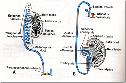
The mesonephric duct elongates and highly coiled to form the:
- epididymis
- vas deferens
- seminal vesicle
- ejaculatory duct
The cranial part of the mesonephric duct forms the:
- appendix of the epididymis
The mesonephric tubules forms the efferent ductule of testis.
The paramesonephric duct is obliterated in males.
Summary,
- Mesonephric duct: Male
- Paramesonephric duct: Female
The mesonephric duct incorporates with the urinary bladder forming the internal trigone, therefore the internal trigone is the only part of the urinary bladder which is mesodermal (the others endoderm).
*What is the importance of the internal trigone?
Anatomically: No submucosa
Physiologically: Most stretch receptors are located in the internal trigone, therefore at 150ml of urine collected in the urinary bladder, the stretch receptors are alerted and hence this is the starting point of the micturition reflex.
Anomalies of the urinary bladder/tract
1) Penile abnormalities
Abrnomal fusion of the urethral plates (folds) during the development of penile urethra is the causative factor
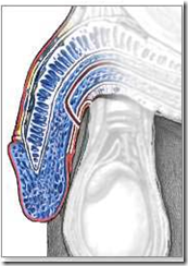
- Epispadius
The abnormality of the urethral development where the urethra generally opens on the dorsum (top/side) of the penis rather than the tip.
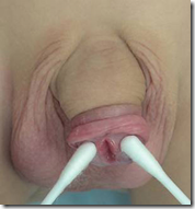
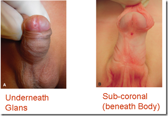
- Hypospadius
Relatively common abnormality in which the opening of the urethra is on the underside of the penis
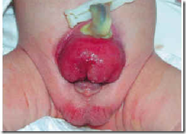
2) Ectopia vesicae/Bladder exstrophy
It is a rare development abnormality that is present at birth.
The bladder & related structures are turned inside out, the posterior vesical wall turns outward (exstrophy) through an opening in the abdominal wall and urine is excreted through this opening. There is deficiency of the anterior abdominal wall.
3) Patent allantois
4) Micropenis
_____________________________________________________________________________________________
Question:
A 2 year old child is brought to the hospital with complaints of difficulty in urination. On examination, the external urethral meatus is found on the under aspect of the penis in the middle (hypospadius). Which of the following developmental error is true for this patient?
- Abnormality of the ureteric bud (kidney)
- Abnormality of the metanephros (kidney)
- Abnormality of the vesico-urethral canal (urinary bladder)
- Abnormality of the urethral plate (CORRECT)
- Abnormality of the mesonephric duct (up til ejaculatory duct only)


