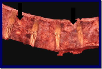
Osteomyelitis
- Definition: Inflammation of the bone & marrow
- Challenging to treat
- less choice of effective antibiotics
- less penetrance of antibiotics into the bone
- infection will lead to disability/morbidity!
- viruses
- bacteria
- parasites
- fungi
2 types of osteomyelitis
- Pyogenic osteomyelitis (Acute)
- Infection reach bone by
- Hematogenous spread (most common)
- Extension from contiguous site
- Direct implantation
- Microorganisms
- Staphylococcus Aureus (most common)
- UTI & IV drug abusers
- E. Coli
- Pseudomonas
- Klebsiella
- Haemophilus influenza
- children
- Salmonella
- sickle cell anemia
- Pathophysiology
- On Location:
- infection reaches metaphysis
- bacteria proliferates
- causing bone death
- Sequestrum: piece of dead bone
- bone necrosis
- causing bone death
- sub-periosteal abscess formation
- lifts the periosteum
- further impair the blood supply
- Rupture of Periosteum
- abscess in surrounding soft tissue
- Sinus formation
- to drain fluid
- Host response:
- Release cytokines from leukocytes
- stimulate osteoclastic bone resorption
- Fibrous tissue formation
- Reactive bone deposition in periphery
- Forms a sleeve of new viable bone
- Involucrum: new bone
- Release cytokines from leukocytes
- On Location:
- Clinical course
- Acute systemic illness
- Pain
- Tenderness
- Immobility
- Xray
- Lytic focus of bone destruction
- Followed by zone of sclerosis
- Blood culture
- can be +ve
- Biopsy
- invasive
- confirms the diagnosis
- Acute systemic illness
- Complications
- Pathological fracture
- Secondary amyloidosis
- Endocarditis
- Sepsis
- Sinus tract squamous cell carcinoma (rare)
- Epiphyseal infection
- Joint cartilage is rigid
- last to get infected
- infection spreads to articular surface
- spreads to joint capsule
- suppurative/septic arthritis
- lead to extensive destruction of articular cartilage
- permanent disability
- Joint cartilage is rigid
- Infection reach bone by
- Mycobacterial/Tuberculous osteomyelitis (Chronic)
- 1-3% of TB cases
- Infection sites:
- Ribs
- direct involvement
- Vertebrae
- from lymphatic drainage
- common in thoracic & lumbar vertebrae
- Ribs
- Spine TB: Pott’s spine
- Infection breaks through intervertebral disc
- involve vertebrae & soft tissue
- forming abscesses
- Infection breaks through intervertebral disc
- Complications
- Psoas abscess formation
- Compression fracture
- of the vertebrae
- Scoliosis or kyphosis
- Spinal cord compression
- neurological deficits
- TB arthritis
- Abscess
- Sinus tract formation
_____________________________________________________________
Bone tumours
- Rare
- 40% malignant
- Aetiology
- No obvious cause (usually)
- Ionising radiation
- Predisposing conditions:
- Paget’s disease
- Fibrous dysplasia
- Retinoblastoma
- genetic
- Syndromes
- Gardner’s
- Ollier’s disease
Category/Classification
- Primary
- Bone-producing tumours
- Osteoid osteoma
- Osteoblastoma
- paediatric
- Osteosarcoma*
- Cartilage-producing tumours
- Osteochondroma*
- Chondroma (enchondroma)
- Chondrosarcoma*
- Miscellaneous tumors
- Ewing’s sarcoma*
- Giant cell tumor of bone*
- Secondary
- Metastases
- Tumour-like bone tumour (mimicry)
- Bone cysts
- Simple bone cyst
- Aneurysmal bone cyst
- Fibrous-osseous lesion
- Fibrous dysplasia
- Eosinophilic granuloma
- Langerhans histiocytosis
_____________________________________________________________________
Primary (more common in the young)
Osteosarcoma

Codman’s triangle & Sunburst appearance in Osteosarcoma
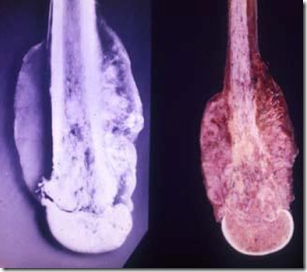

Osteosarcoma – spread towards cortex
- Because bone is a CT
- most common primary tumour
- young adults (10-25 yo)
- rare in later age, due to secondary causes
- Genetic
- mutations to RG gene
- Morphology
- Location
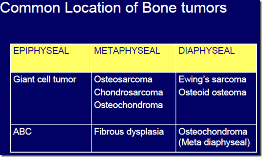
- metaphysis of long bone
- around knee
- tibia, fibula
- In old age
- flat & long bones
- Anatomical portion of bone
- Intramedullary
- most common
- tumour can break through cortex
- Intracortical
- Surface
- good prognosis
- Gross
- Form bulky tumours
- Gritty, gray white, yellowish mass
- Invasion to surrounding tissues
- Infiltrate the medullary canal & spread towards cortex
- epiphyseal penetration is rare
- Joint invasion
- later stages
- Microscopy
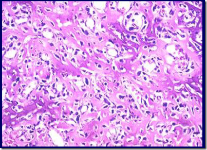
- Tumour cells are osteoblastic cells
- histologic variants:
- osteoblastic
- chrondroblastic
- small cell
- Formation of lacy osteoid by tumour cells
- Variation in size, shape
- bizarre features
- Tumour giant cells
- Hyperchromatism
- Mitiosis
- Binucleation
- Vascular & blood vessels invasion
- Xray
- large destructive mass lesion
- mixed lytic & sclerotic areas
- Elevation of periosteum
- Codman’s triangle
- Sunburst appearance
- spread of tumour into surrounding soft tissue
_____________________________________________________________________
Osteochondroma
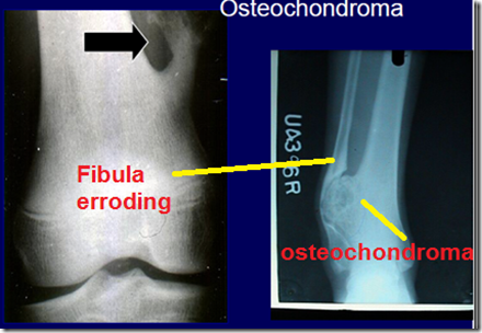
- Cartilage covered by bony excrescense (exostosis)
- formation of new bone on surface of bone
- Can be solitary or multiple
- Spontaneous
- Location
- metaphysis
- diaphysis
- Morphology
- painless skeletal swelling
- slowly growing mass
- palpable bony masses
- Complications
- can lead to limb shortening
- when bone is still actively growing
- 1st – 2nd decade of life
- fractures
- bone deformities
- neurologic/vascular injuries
- bursa formation
- malignant transformation
- rare
- Osteochondromatosis
- autosomal dominant condition
- short stature
- multiple osteochondroma
- located close to metaphysis
- sessile or pedunculated
- cortex of lesion continuous with cortex of bone
- with a homogenous continuation of the medulla
- asymmetric growth
- knees
- ankles
- may lead to deformities
_____________________________________________________________________
Chondrosarcoma

Chondrosarcoma – affects axial skeleton

Chondrosarcoma – affecting the finger
- 2nd most common malignant primary bone tumour
- older adults
- 30-60 yo
- can arise from previous enchondroma
- many are sporadic
- location
- axial skeleton
- pelvis
- pectoral girdles
- ribs
- spine
- Aggressive
- erodes & invades soft tissue
- *Metastasize to: (suspect chondrosarcoma)
- lungs
- liver
- kidney
- brain
- Gross
- large bulky tumours
- grey white, translucent
- Microscopy
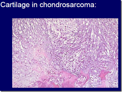
- malignant cartilage
- with anaplastic chondrocytes in spaces
- focal enchondral ossification & calcification
- hypercellularity, atypia, mitosis
- Treatment
- resistant to chemotherapy
- therefore must be surgical resection
_____________________________________________________________________
Giant cell tumour of bone (Osteoclastoma)
- Osteoclastoma
- potentially malignant
- mostly benign
- arise in epiphysis
- develop after fusion of cartilage
- after epiphyseal plate thins out
- lytic lesion at end of bone
- knee
- elbow
- ankle
- Treatment
- resection
- recurrence is common
- Pathology
- osteoclastic giant cells
- 100 or more nuclei
- nuclei similar to those of stromal cells
- giant cells are not malignant
- mesenchymal cells
- ovoid
- mononuclear cells
- Haemorrhage and necrosis present
_____________________________________________________________________
Secondary (metastatic)
 Looking at the pattern of bone lesion, you should also screen for these primary cancer sites
- more common in the old
- most commmon malignant tumour in skeleton
- metastasize from:
- breast tumour
- kidney tumour
- thyroid
- lung
- Generally occur in
- vertebrae
- pelvis
- if primary tumour from pelvis, can spread to lumbosacral spine
- proximal parts of the
- femur
- humerus
- ribs
- skull
- Rare
- hands
- feet
- Marrow neoplasm (hemopoietic):
- myeloma
- leukemia
- lymphoma
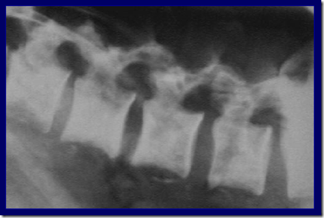
Mixed lytic and sclerotic lesion


