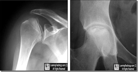
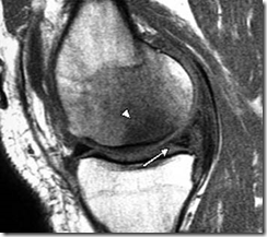
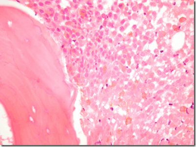
Types of bone disorders:
- Inherited
- Achondroplasia
- Osteogenesis Imperfecta
- Osteopetrosis
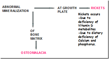
- Abnormal mineral homeostasis/mineralization
- Rickets
- Osteomalacia
- Renal osteodystrophy
- Abnormal matrix
- Osteoporosis
- Osteogenesis Imperfecta
- Reduced bone mass
- Neoplasia
- multiple myeloma
- carcinomatosis
- Osteoclast dysfunction
- Osteopetrosis
- Pagets disease of the bone
- Caused by renal dysfunction
- Renal osteodystrophy
- Circulatory disturbances
- Osteonecrosis (Avascular necrosis)
_____________________________________________________________________
Inherited bone disorders
Achondroplasia
- common form of short limb dwarfism
- autosomal dominant trait
- mutations in fibroblast growth factor receptor-3 (FGFR3) gene
- Features
- disproportionate short stature
- shortening of arms and legs
- normal trunk length
- megalencephaly
- unusually large brain
- frontal bossing
- midface hypoplasia
Abnormal mineral homeostasis/mineralization
Normal mineralization requires adequate calcium & phosphate.
Calcitriol (Activated Vit D/DHCC)
Vitamin D metabolism:
Role of calcitriol/DHCC/Vit D:
- promotes absorption of Ca & Phosphate from intestine
- increases reabsorption of P04 in the kidney
- acts on bone to release Ca & PO4
- increases conc of CA & PO4 in the ECF
- leads to
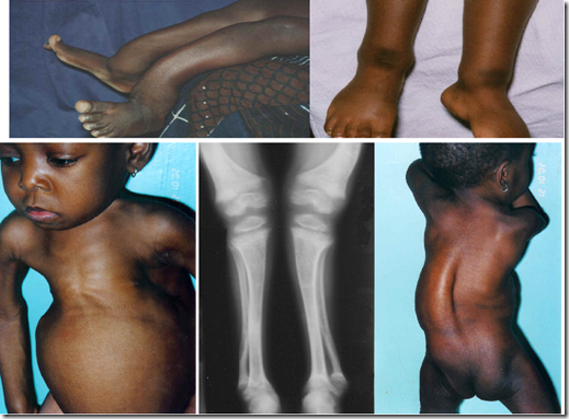
Rickets
- Predisposing causes
- Vitamin D deficiency
- Dietary
- Reduced sunlight
- Dark skin
- Malabsorption
- small intestine diseases
- gastrectomy
- hepatobiliary disease
- chronic pancreatic insufficiency
- Disorders of Vitamin D metabolism
- use of anticonvulsants
- Renal
- Chronic renal failure
- Distal renal tubular acidosis
- Secondary renal acidosis
- Drug induced
- PB
- tetracycline
- CRF
- transplants
- Phosphate depletion
- dietary (low PO4 intake)
- antacids
- hereditary
- hypophosphatemic rickets/osteomalacia
- acquired
- tumour-associated rickets
- osteomalacia
- states of rapid bone formation (with or without a relative defect with bone resorption)
- postoperative hyperparathyroidism
- osteopetrosis
- Pathology
- overgrowth of epiphyseal cartilage
- due to insufficient calcification
- failure of cartilage cells to mature and disintegrate
- deposition of organic matrix on inadequately mineralized caltilage
- enlargement and lateral expansion of costochondral junction
- Abnormal overgrowth of capillaries & fibroblasts
- due to microfractures in poorly formed bones
- Pathological features
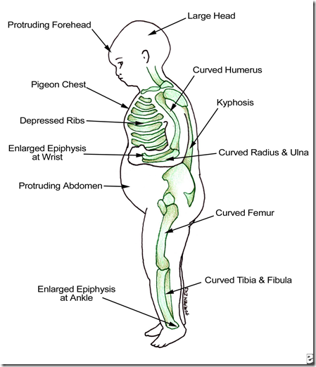
- Skull
- Craniotabes
- bone of skull soften
- flattening of the posterior skull seen
- Frontal bossing
- sometimes square forehead
- caput quadratum
- Teeth
- Erupt later than normal
- Enamel of poor quality
- dental caries
- Thorax
- Rickety rosary
- enlarged end of ribs resembling beads
- can be palpable and visible at costochondral junction
- sternum become more prominent
- pectus carnatum appearance
- Harrison groove
- Semicoronal appearance over the abdomen
- at the diaphragm level
- Spine
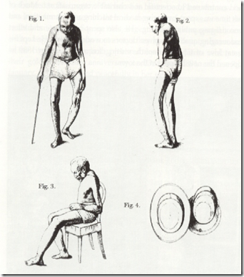
- Mild to moderate pronounced scoliosis
- Pelvis
- Prominant promontary
- AP diameter of pelvis shrink
- due to scoliosis
- in girls, complication during childbirth
- Arms
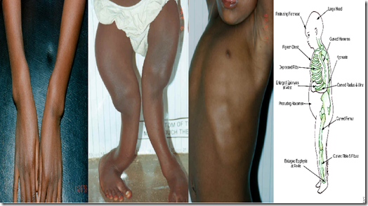
- Bowing of long bones
- reaction to greenstick fractures
- fracture in a young, soft bone in which the bone bends and partially breaks
- as a result of concurrent osteomalacia
- Thickening of wrist epiphysis
- fraying and cupping of metaphysis
- Legs
- Bowing of long bones
- genu varum
- due to weight bearing
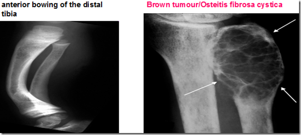
- anterior bowing of tibia
- saber shin deformity
- Development of knock knees
- genu valgum
- due to displacement of growth plates
- Laxity/looseness in the ligaments
- Decreased muscle tone
- Delay in motor development
Osteomalacia
- Equivalent to rickets
- but in adults
- milder form
- Pathology
- Inadequate mineralization of bone
- following cessation of bone growth
- Laboratory investigation
- Blood serum
- Ca low
- PO4 low
- Alkaline phosphatase high
- PTH high
- X-ray
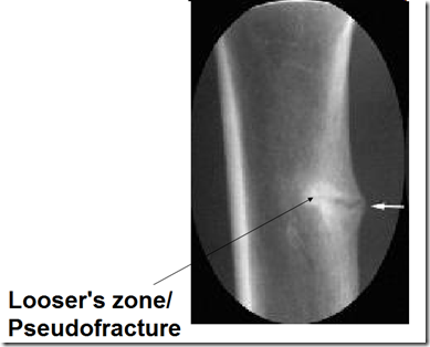
- looser’s zones in xray
- pseudofractures
- Protrusio acetabuli
- hip joint disorder
- Microscopy
- Aetiology
- Vitamin D deficiency
- dietary
- low sunlight exposure
- malabsorption
- Abnormal Vitamin D metabolism
- liver disease
- renal disease
- Drugs
- anticonvulsant
- Hypophosphatasia
- dietary
- genetic
- Clinical manifestation
- malaise
- bone pain
- proximal muscle weakness
- Treatment
- Oral Vitamin D replacement
_____________________________________________________________
Abnormal Matrix
Osteogenesis Imperfecta
- Cause
- deficiency in synthesis of type 1 collagen
- mutation in genes
- COL 1A1 & COL 1A2
- Basic abnormality
- too little bone
- osteoporosis with marked cortical thinning
- attenuation of trabeculae
- Anatomical structure rich in type 1 collagen affected
- joints
- eyes
- ears
- skin
- teeth
- Investigations
- Collagen synthesis analysis
- by culturing dermal fibroblasts
- obtained during skin biopsy
- Prenatal DNA mutation analysis
- analyze uncultured chorionic villus cells
- Types
- 1
- mild
- compatible with survival
- autosomal dominant
- Decreased/abnormal synthesis of pro-α1(1) chain
- Clinical presentation
- postnatal fractures
- skeletal fragility
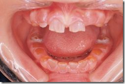
- dentinogenesis imperfecta
- teeth discoloured/translucent
- weaker than normal
- hearing impairment
- joint laxity/looseness
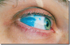
- blue sclerae
- sclera of the eyes are blue
- 2
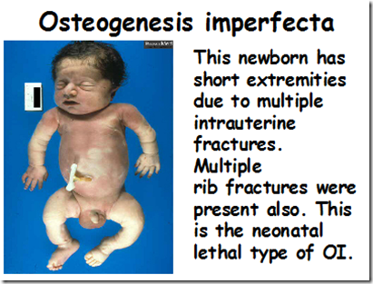
- also known as neonatal lethal type of Osteogenesis Imperfecta
- extremely severe
- most lethal in perinatal
- death in utero/within days after birth
- autosomal recessive
- Abnormal or insufficient pro-α2
- Clinical presentation
- Skeletal deformity with excessive fragility and multiple fractures
- Blue sclerae
- 3
- severe
- autosomal dominant
- 4
- undefined
Osteoporosis

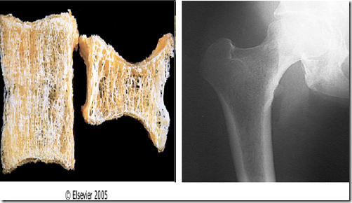
Comparing normal microarchitecture of bone to an osteoporotic one.
- Most common metabolic bone disorders
- Characterised by
- reduced bone mass
- microarchitectural detrioration of bone tissue
- increased bone fragility
- susceptibility in fracture
- Pathophysiology
- bone remodelling occurs throughout our lifetime
- In normal adults,
- activity of osteoclasts balanced by activity of osteoblasts
- With onset of menopause
- diminished oestrogen
- exc
essive bone resorption - not fully compensated with bone formation
- Why do we have to recognise osteoporosis
- To prevent fractures & its complications
- Types
- Type 1 (postmenopausal)
- oestrogen/testosterone deficiency
- regardless of age
- bone resorption outpaces bone formation
- decrease in trabeculae bone
- increase risks of fractures
- Type 2
- occurs to both men & women
- Reduced bone formation
- loss of cortical & trabeculae bone
- increased risk fractures
- Decreased renal production of calcitriol (activated vitamin D) occurring late in life
- Type 3
- Secondary osteoporosis, due to:
- medication
- glucocorticoids
- diseases
- hypogonadism
- hypercortisolemia
- hyperthyroidism
- hyperparathyroidism
- anorexia
- renal failure
- chronic liver disease
- malabsorption
- celiac sprue
- surgical
- inflammatory bowel disease
- pregnancy
- type 1 diabetes
- HIV
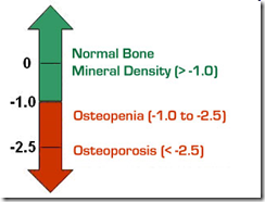
- WHO criteria for diagnosis
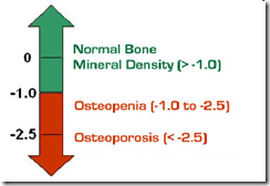
- T-score
- <-1
- normal
- low risk
- check again in few years
- -1 to –2.5
- osteopenia (low bone mass)
- moderate risk
- treat if have other risk factors
- -2.5 or greater
- osteopororsis
- high risk
- treat
- –2.5 or greater + fx(s)
- severe/established osteoporosis
- high risk
- treat
- Management
- Prevent first fragility fracture/future fractures
- initiate lifestyle changes
- Relieve symptoms of fractures/skeletal deformities
- Stabilize/increase bone mass
- Improve mobility/functional status
- Medication
- Hormone replacement therapy (HRT)
- Raloxifene
- oral selective estrogen receptor modulator (SERM) that has estrogenic actions on bone
- Calcitonin
- Biphosphonates
- a class of drugs that prevent the loss of bone mass
- PTH
OLIS: http://elearning.imu.edu.my/file.php/4236/StudyGuide/Musculo/week1/xms1_5.htm
_____________________________________________________________________
Reduced bone mass (due to neoplasia)
- Causes
- Neoplasia
- Multiple myeloma
- Carcinomatosis
- cytokines produced by malignant cells activates osteoclasts
- Alcohol
- direct effect on bones
- Immobilization
- bone loss
- Pulmonary disease
- Homocystinuria
- Smoking
- increased hepatic metabolism of oestrogen
- direct effect on bone
- earlier menopause
- Medications
- corticosteroids
- inhibit bone formation
- inhibit Ca absorption
- heparin (high dose)
- Aluminum
- Anticonvulsants
- phenobarbitol
- phenytoin
- induce vitamin D deficiency
- reduce intestinal Ca absorption (?)
- Cyclosporin
- Aromatase inhibitors
- Antiretroviral therapy
- Clinical features
- Fractures
- mid to lower thoracic / upper spine
- verterbral fractures
- acute back pain after bending
- lifting
- coughing
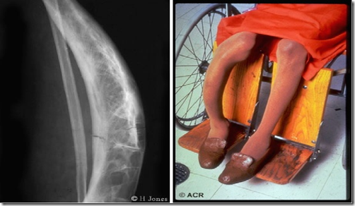
- Progressive kyphosis with loss of height
- Pain
- sharp, nagging or dull
- sometimes radiate to abdomen
_____________________________________________________________________
Osteoclast dysfunction
Osteopetrosis
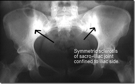
Diffuse symmetric skeletal sclerosis
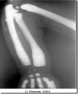
Bone sclerosis is a condition in which bones thicken, harden or increase in density.
- Also known as:
- marble bone disease
- Albers-Schönberg disease
- Osteopetrosis, term coined because:
- stone-like quality of bone
- bone extremely brittle
- fracture like a piece of chalk
- A group of rare genetic disorders
- Characterized by:
- reduced osteoclast bone resorption
- resulting in diffuse symmetric skeletal sclerosis
- sclerosis seen as bright areas in xray
- bones lack a medullary canal
- usually formed via remodelling process
- bones formed not remodelled
- Erlenmeyer flask deformity
- ends of long bones are bulbous and misshapen
- metaphysis
- Neural foramina becomes smaller
- compress exiting nerves
- The foramen allows for the passage of
the spinal nerve root, dorsal root ganglion, the spinal artery of the segmental artery, communicating veins between the internal and external plexuses, recurrent meningeal nerves, and transforaminal ligaments. - Classified into variants (based on mode of inheritance and clinical features)
- Autosomal recessive malignant type
- Autosomal dominant benign type
Osteitis Deformans (Paget’s disease of the bone)
- Most common bone disorder (after osteoporosis) in elderly
- 3: 2 , Male-female ratio
- Aetiology
- slow virus infection by a paramyxovirus
- Osteoclast precursors hyperresponsive to the RANK ligand (RANKL)
- which promotes osteoclast genesis
- Clinical features
- Old Hat don’t fit anymore!
- hat gets tighter
- head diameter becomes larger
- Bone pain
- most common
- Joint pain
- Deformity
- Spontaneous fracture
- Hypercementosis of teeth
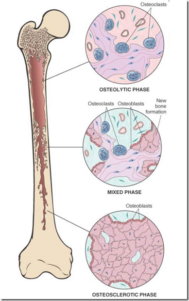
- Pathogenesis
- Initial osteolytic stage
- regions of furious osteoclastic bone resorption
- Mixed osteoclastic-osteoblastic stage
- hectic bone formation
- compensatory reaction
- Burnt-out quiescent osteosclerotic stage
- bone cell activity becomes markedly diminished
- Nett effect
- Gain in bone mass
- however, the newly formed bone is disordered and architecturally unsound
- Investigations
- Serum
- Alkaline phosphatase high
- Ca2+ normal
- Urinary hydroxyproline, pyridinoline cross links
- markers of bone resorption
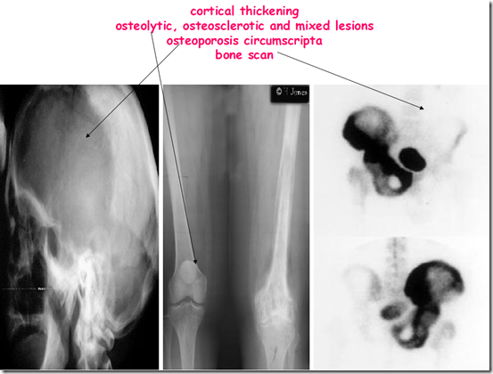
- Xray
- cortical thickening
- osteolytic, osteosclerotic and mixed lesions
- osteoporosis circumscripta
- well-defined lysis, most commonly in frontal bone producing well-defined geographic lytic lesion in skull
- Represents early destructive phase of disease active stage
- Bone biopsy
- Mosaic pattern of lamellar bone pathognomonic(characteristic) of Paget disease.
- larger than normal
- composed of coarsely thickened trabeculae
- cortices that are soft and porous and lack structural stability
- Complications
- Skeletal
- deformity
- fracture
- secondary osteoarthritis
- dental complications
- primary bone tumours
- osteosarcoma
- Skeletal/neurological
- deafness
- Neurological
- cranial nerve palsies
- spinal stenosis
- hydrocephalus
- an abnormal accumulation of cerebral spinal fluid(CSF) in the ventricals/cavities of brain
- Others
- High output cardiac failure
- Immobilisation hypercalcaemia
- cardiac valvular calcification
_____________________________________________________________________
Renal Dysfunction
Renal osteodystrophy
- Pathogenesis
- Due to renal failure, there is retention of phosphates
- resulting in hyperphosphatemia
- which causes hypocalcemia
- due to precipitation of phosphate with the calcium in tissues
- therefore less in serum
- Hypocalcemia will trigger release of PTH
- resulting in secondary hyperparathyroidism
- Hyperparathyroidism
- increases bone resorption
- to increase serum calcium level
- Characteristics (all bone disorders seen)
- Increased osteoclastic bone resorption
- Delayed matrix mineralisation
- osteomalacia
- Osteosclerosis
- increased proportion of cancellous bone where calcium may be deposited as amorphous calcium phosphate rather than hydroxyapatite
- Osteoporosis
- Growth retardation
- Rickets
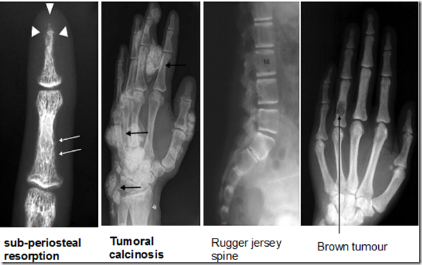
- Features of hyperparathyroidism
- Diagnosis
_____________________________________________________________________
Circulatory Disturbances
Osteonecrosis
- Also known as:
- Aseptic bone necrosis
- Avascular bone necrosis
- Bone infaction
- Most often affects children & adolescents
- because growing bone requires more oxygen than adult bones
- May occur in any bone, but most important sites:
- carpal bones
- head of femur
- Caused by
- inadequate blood supply to the bone
- by trauma or damage to blood vessels
- vasculitis
- inflammation to the blood vessels
- sickle cell anemia
- alcohol abuse
- fat embolism
- trauma, causing blockage
- hypercoagulable states
- systemic diseases
- gout
- artherosclerosis
- diabetes mellitus
- Clinical features
- Painful restricted movements of joints
- tenderness
- muscle wasting
- Osteonecrosis is followed by a regenerative process in surrounding tissues
- increased osteoclastic activity to remove necrotic bone
- increased osteoblastic activity – repair
- Treatment
- Total hip arthoplasty
- joints
- Core decompression
- Bone graft


