General overview
- Ascending tracts
- Sensory
- Descending tracts
- Motor
General arrangement of both tracts
- 1st order neuron
- 2nd order neuron
- 3rd order neuron
The only difference is the different locations where each order of neuron ends.
Decussation is the cross-over of the tract from one side to the other. Therefore, there are instances where the left side of the body is controlled by the right brain hemisphere. Decussation occurs at different locations for each tracts.
_____________________________________________________________________
Descending tracts (Motor)
General arrangement of descending tracts
- 1st order neuron
- First-order neurons conduct impulses from receptors of the skin and from proprioceptors (receptors located in a join, muscle or tendon) to the spinal cord or brain stem, where they synapse with second-order neurons. First-order neuron’s cell bodes reside in ganglion (dorsal root or cranial).
- starts at the cerebral cortex in the somatomotor area
- 2nd order neuron
- 2nd neuron to carry an order. The order could be a sensory stimulus or a motor stimulus.
- axon of the 1st order neuron will synapse with the 2nd order neuron at the level of the brain stem, which commonly decussate (crosses over) to the opposite side.
- 3rd order neuron
- The 3rd order neuron is located in the ventral horn of the spinal cord, which will exit with the spinal nerve to supply the muscle.
Types of descending tracts:
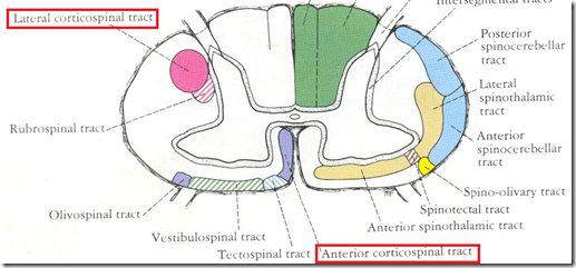
- Lateral corticospinal tract
- Anterior corticospinal tract
Therefore, the descending tract is also known as corticospinal tract.
Flow of arrangement of the corticospinal tract:
- Corticospinal tract arise from
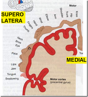
- long axons of the pyramidal cells (extrapyramidal layer) of the precentral gyrus (primary motor centre of the cerebral cortex)
- lies in front of the central sulcus
- Homunculus arrangement
- arranged upside down
- the finer the movement, the more the cortical representation
- fingers, face, tongue – more
- trunk, lower limbs – less
- medial surface: lower limbs
- superolateral surface: everything else
1) 1st order neuron
- Fibres of the 1st order neuron arise from the precentral gyrus
- These fibres converge and enter a small area
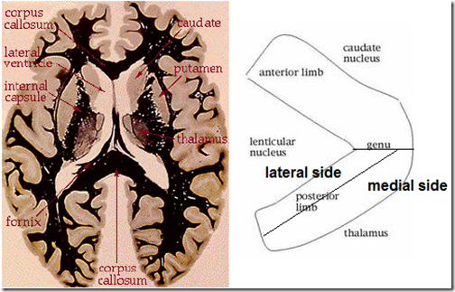
- internal capsule
- like a bunch of flowers with a ribbon tied around it
- ALL the fibers (from ascending & descending tracts) converge here
- Function: separates the caudate nucleus and the thalamus from the lenticular nucleus
- Ques: location of internal capsule
- bounded medially by the thalamus and caudate nucleus
- bounded laterally by the lenticular nucleus
- Parts of internal capsule (not homunculus arrangement, normal head to toe)
- anterior limb
- head & neck fibres most anterior
- posterior limb
- lower limb fibres most posterior
- The descending fibres passes through the
- LATERAL half of the posterior limb of internal capsule
- After the internal capsule, the fibres enter the brain stem
- midbrain
- pons
- medulla
2) 2nd order neuron
- Fibres of the 1st order neuron ends when it enters the brain stem and synapse with the 2nd order neuron
- The fibres pass through the brainstem
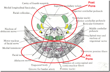
- 1st – through the (mid 5th) crus cerebri of midbrain
- 2nd – through the anterior part of the pons
- 3rd – in the medulla oblongata
- 80-85% of the fibres cross to the opposite side
- Motor decussation
- uncrossed fibres
- Enters the spinal cord
3) 3rd order neuron
- 2nd order neuron fibres in the medulla oblongata enters the spinal cord and synapse with the 3rd order neuron
- Motor decussation
- in the spinal tract, the crossed tract descend as the lateral corticospinal tract
- Therefore, the motor cortex of the cerebral hemisphere controls the opposite side of the body (L – R, R – L)
- contra-lateral side
- In upper motor neuron lesions:
- above the motor decussation (above medulla)
- opposite side of body affected
- below the motor decussation
- same side of body affected
- ipsilateral side
- Uncrossed fibres
- in the spinal tract, the uncrossed tract descent as the anterior corticospinal tract
- its fibres cross at spinal level?
_____________________________________________________________________
Ascending tracts (sensory)
Types of ascending tracts:
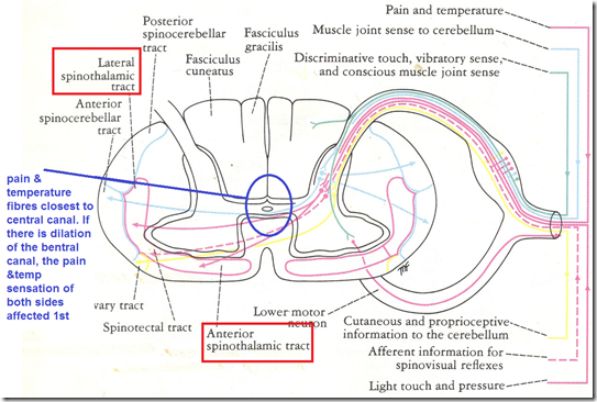
- Spinothalamic tracts
- Lateral
- pain & temperature
- Anterior
- light touch & pressure
- Dorsal column tract
- deep touch & pressure
- proprioception
- vibration sensation
- Spinocerrebellar tract
- posture & coordination
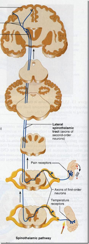
Flow of arrangement of Spinothalamic tracts:
1st order neuron:
- Arise from sensory receptors of the body
- The fibres enter the white mater and ends at the substantia gelatinosa
- tip of posterior gray horn
2nd order neuron:
- The fibres of 1st order neuron synapse with the 2nd order neuron at the substantia gelatinosa
- These fibres then cross to the opposite side
- Pain & temperature fibres
- enters the lateral spinothalamic tract
- Light touch & pressure fibres
- enters the anterior spinothalamic tract
- These tracts ascends to brainstem
- to medulla oblongata, pons and midbrain
- tracts flattened in the brainstem
- spinal lemniscus
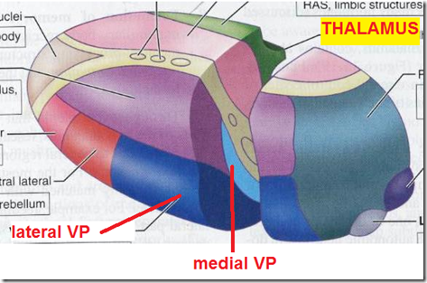
- Reaches the ventral posterolateral nucleus of the thalamus
- ends here
3rd order neuron:
- The 3rd order neurons arise from the thalamus and pass through the internal capsule
- thalamocortical fibres pass through the medial part of the posterior limb of the internal capsule
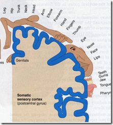
- Enters the postcentral gyrus
- sensory cortex of the cerebrum
- behind the central sulcus
- Same homunculus arrangement
- more sensitive areas in the body have a greater representation
_____________________________________________________________________
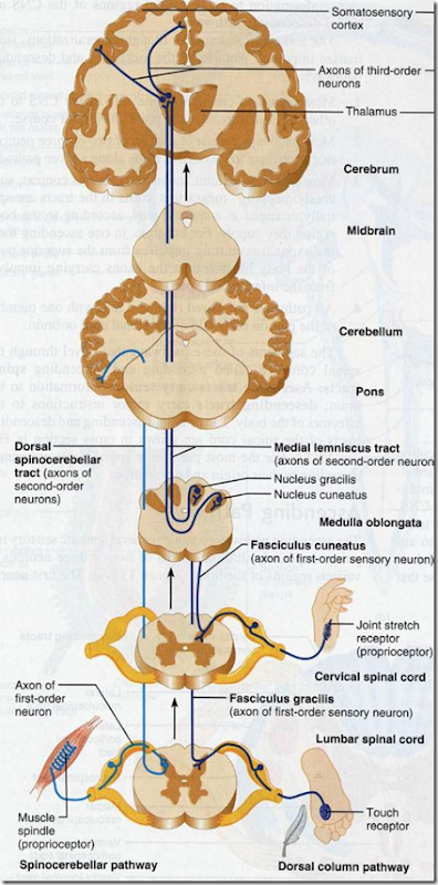
Flow of arrangement of the dorsal column tracts:
1st order neuron:
- Arise from the sensory receptors of the body
- Fibres enter the dorsal column of the SAME side (post column of spinal cord)
- ascends to the medulla oblongata
- (does not synapse and end here like spinothalamic tract)
- Enters medulla oblongata
- ends in the gracile and cuneate nucleus
2nd order neuron:
- Starts at the gracile & cuneate nucleus of the medulla oblongata
- These fibres crosses to the opposite side of the medulla oblongata
- Ascends through the brain stem
- as flattened bundle
- medial lemniscus
- Ends in the ventral posterolateral nucleus of the thalamus
3rd order of nucleus:
- Arise from the thalamus
- Pass through the internal capsule
- medial aspect of the posterior limb of internal capsule
- Reaches the postcentral gyrus
- ends here
_____________________________________________________________________
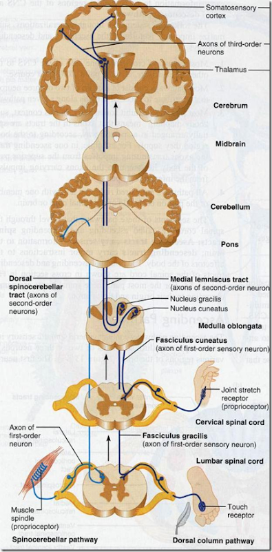
Flow of arrangement of Spinocerebellar tract:
1st order neurons:
- Arise from the sensory receptors of the body
- Enters the spinal cord
- Ends in the Clarke’s Column of the posterior grey horn
- synapse
2nd order neurons:
- Arise from the Clarke’s Column
- synapse with 1st order neurons
- Ascends in the spinocerebellar tracts
- Enters the cerebellum
- through the interior and superior cerebellar peduncles
- the only tract that enters the cerebellum
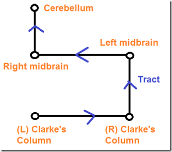
Actual decussation of these tracts:
- These tracts decussate 2 times
- therefore cerebellum controls same side of body
- ipsilateral
- eg. right spinocerebellar tract controls the right side
- vice versa
_____________________________________________________________________
Clinical anatomy:
- Lesion of one half of the spinal cord may lead to
- Opposite side of body
- loss of pain, temperature, light touch, pressure sensations
- Same side of body
- loss of the other sensations
- Sensory cortex of the cerebral hemisphere controls the opposite side of th
e body - contralateral
- Lesion above the sensory decussation
- all the sensations of the OPPOSITE side of the body are lost
- Lesion below the sensory decussation
- sensations of the SAME side of the body are lost
- Cortical lesions
- affected areas are usually limited
- paralysis/parasthesia is localized
- Internal capsule lesions
- all ascending & descending tracts are affected
- hemiplegia/hemiparasthesia





useful and clear.
Thanks a lot
Glad to be of help!
You’re welcome!
Not everything is clear. Too much summary results in vagueness; words without meaning sound with sense.
This has cleared up what lecture notes and wikipedia and textbooks couldnt!! thankyou so so much!! 🙂
You’re welcome! That’s why I started this blog! 🙂
wonderful collection
Sharam kar
thanks a lot for this lecture notes. it helps me a lot. =)
hope to share more notes from this page…thank you….^_^
You’re welcome Syuhada : )
Nice review
Thank you! This has proved very helpful for a PBL session. 🙂
You’re most welcome!
superb clarity
IT WAS GREAT AND USEFUL BUT I COULDNT UNDERSTAND THE SPINOCEREBELLAR TRACT ESP THE LAST DIAGRAM HOW IT MEETS THE RIGHT N LEFT MID BRAIN AS MID BRAIN LIES ABOVE THE CEREBELLAM
This is kind of late but here goes; it is a 2D image think of it in 3D. That’s why it enters the cerebellum through the anterior and superior peduncles.
“internal capsule
Function: separates the caudate nucleus and the thalamus from the lenticular nucleus”
“heart
Function: separates the left lung from the right lung”
7/10 would read again
Think of it in terms of conductivity
Oh well not all points were vague. For maximum benefit use with some other site. gives you a guide.
the theory was quite good but can it be given in little details please for easy understanding..thank you so much for the information….
Am greatful for this note, it of helped to me. Thanks.
Can you explain how the cerellar tract crosses twice? is that the ventral/anterior tract? Have you diagrams of the other ascending/decending tracts? Many many thanks
Ventral spinocerebellar tracts cross immediately they enter spinal cord and again in the white matter of the cerebellum(crossing a road twice) dorsal spinocerebellar tracts don’t decussate at all.
meant cerebellar
Specific functions
hmmm! At least i can now to some xtend the ascending and descending tracts of the spinal cord. Tanks alot
OH THANKS
I love you. None of my textbooks have anything about any tracts in them!! Useless. And wikipedia was a bit useless too! This is amazing. Thank you so much!
An interestin piece. Thanxx a lot!
it is little bt helpful…………
This was very helpfull thankyou
Thank you so much!
Thank you, now can we just have you as our teacher and we will be fine!
Thanks to your notes now I understand what “cross” means. Keep up the good work.
It was a superb review. Thanks.
explained in a simple and interesting way
all concepts cleared…. thanks a lot….
Whoa! There is a massive mistake in this article! In the section titled: “General arrangement of descending tracts” it says that first order neurons collect impulses from the skin and proprioceptors”. The problem is that you’re describing ascending tracts! First order descending tracts start in the brain, not in the periphery. If they start in the skin, or proprioceptors and go to the brain, they would be ASCENDING neurons. Motor neurons start in the brain and DESCEND to the muscles and organs. The funny thing is, the diagram that you used directly above the text is accurate, it shows the first order neuron in the motor cortex. That’s all I’ve read so far.
thankquuuuuuuuuu soooooooooooooooooooo much
Help full for my presentation thanksssssss too
Good forum but I have a question. According to my reading I have seen that the corticospinal tracts (which are descending) consist of only 2 neurons (Upper and Lower) and 1 synapse (in the anterior horn of the spinal column) as compared to the ascending sensory tracts which contain 3 neurons and 2 synapses. My question lies in the above explanation which say that there are 3 neurons in the corticospinal tracts with 2 synapses.
Anyone can clarify?
Really it is one of the best study materials of ascending and descending tract
Thank u so much for this
Very good effort.
thanks!
it is really helpful.
it is very helpful….wish u could cover other topics also
Really helped me a lot! I struggled for a year with the tracts, now it’s so much clearer. Thank you! But just to clarify, for the spinocerebellar pathway , it crosses from the left clarke’s column to the right then up the spinocerebellar tract to the RIGHT mid brain then crosses to the left midbrain to the cerebellum.
woow great, I found it too. and there are spinocerebellar which have not clarified much. and you can share skills too if you are aware of them. thank you
tatenda ………thank you
Reblogged this on kingshamii.
could you please write more on cerebellar tracts?it would help me understand without going through the boring text.
really helped me a lot! thank you so much
The article made me clear of tracts.thnx and gratitude for article..
awesome work
its very easy for understanding thanks a lot
Fantastic it just solved my prob good job
it was real helpful to me in my studies,thanks..!
Thank you its of real good help
I would like some clarification on pathway,vs, faniculus vs, column, vs tract
Wow….amazingly written….simple yet informative…Thanx a lot for taking the effort to summarize and also sharing with everyone!!
thanku so much its really helpful for me
very nice and clear!
Thank you!
nicely summarized
Thanks fr the person. It was useful
fantastic notes they are useful
Thanks
Very good
Very good .I kindly request you to send this study materials to my e mail address
there are a lot of mistakes in it.plz correct them.
Hi Sravya, will be great if you can indicate the mistakes & also the right answers. It will help everyone looking for answers. Thanks
thanks so much for the lectures
thank you so much for sharing your knowledge to all……. regards from inida
very nice and self explanatory
There’s no such thing as a third order neuron for the corticospinal tracts. Nor do the second order neurons stop at the brainstem for corticopspinal tract.
Sensory/Ascending pathway information is fine, but descending/motor pathway is completely wrong.
It solves a lot of questions., Thanks a lot
Hi! What book did you get the images from because I`m searching for pictures like these for all existing tracts! Thanks a lot
thanks
Am really grateful to have gone through this piece of work. It has broaden my scope about Ascending and descending tract of the spinal cord. Thank you very much
God bless you