
Prenatal Development of Central Nervous System

At the beginning of the 3rd week, developing notochord induces overlying ectoderm to become neuroectoderm. Thus forming an elongated, slipper-shaped plate of thickened ectoderm – the neural plate.
- Neural plate invaginates and forms neural tube
- Pre-requirement for folding
- Notochord
- Anterior part
- Forms
- Forebrain
- Midbrain
- Hindbrain

This neural plate invaginates, and the lateral edges of the plate becomes elevated to form the neural folds. This depressed region is the neural groove.
With further development, the neural folds become more elevated and each end fuses at the dorsal midline to form the neural tube.
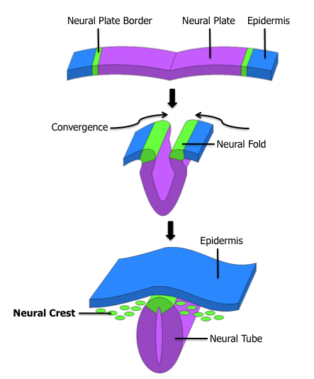
After the fusion, the non neural ectoderm forms the epidermis. At the neural plate border where the neural plate converges, are the neural crest cells. The neural crest cells undergoes a epithelial to messenchymal transition, delaminating from the neuroepithelium and migrating through the periphery where they differentiate into many cell types.
- Therefore, neural crest forms the PNS.
- Structures derived from neural crest:
- Dorsal root ganglia
- Schwann Cells
- Melanocytes
- Pia-arachnoid (Leptomeninges)
- Autonomic neurons
- Adrenal medulla
- During this time, susceptible to Fetal alcohol syndrome.
- neural crest cells affected by ethanol
- many mental/physical disability can arise from this

- The fusion of neural plate begins from the 4th somite (cervical region)
- There are 2 parts where the neural tube is still open (no fusion yet):
- Cranial neuropore
- Caudal neuropore
- These opened parts will close only at
- 25th – 27th day of life (1st month after birth)
_____________________________________________________________________
Layers of neural tube

- Neural tube is composed of 3 layers
- Neuroepithelial layer (neuroblasts)
- innermost
- It is a thick layer of pseudo-stratified epithelium lining the lumen of the neural tube.
- Its component cells undergo rapid cell division to form new cells – neuroblasts
- divide into – apolar –> bipolar –> multipolar
- which migrates to the adjacent mantle zone
- Forms:
- Gliablasts
- astrocytes
- oligodendrocytes
- Mesenchymal cells
- Microglial cells
- Mantle layer (gray mater)
- middle
- The neuroblasts migrated from the neuroepithelium forms a zone around the neuroepithelial layer
- The neuroblasts are characterized by
- large round nuclues
- pale nucleoplasm
- dark staining nucleolus
- Mantle zone later forms the gray mater of spinal cord
- Marginal layer (white mater)
- outermost
- predominantly composed of nerve fibers (processes of cells in the mantle zone)
- Marginal zone later forms the white mater of the spinal cord.
_____________________________________________________________
6 stages of neural cell development
Prenatal Cellular development in brain

- Stage 1
- Mitosis/ Neurogenesis
- neuroepithelial cell –> form neuroblasts
- in the ventricular zone
- Stage 2
- Migration
- from ventricular zone to their destination
- migration aided by glial cells
- acts like ladder
- Filopodia assist in finding location after leaving radial glial cells
- Abnormal migration will lead to
- learning disabilities
- schizophrenia
- autism
- Stage 3
- Differentiation
- gives rise to specific neurons & glial cells
- Stage 4
- Synaptogenesis
- forming synapse with other neurons
- dictates intelligence of fetus
- Involved:
- Neuronal maturation
- Elongation of axons
- Established terminals
- Elongation of dendrites – form new spines
- Expression of neurotrophins (from dendrites)
- Neurotrophic factors
- stimulate cell growth
- helps neurons to mature
- Stage 5
- Cell death
- Apoptosis – active cell death during development
- Necrosis – passive cell death due to injury
- Stage 6
- Synapse rearrangement
- depends on cognitive input & apoptosis


_____________________________________________________________________
Primary & Secondary brain vesicles


_____________________________________________________________________
Post-natal brain development

- At birth, brain weights 25% of full adult brain
- Growth spurt of brain up til 4th year
- At age 6, increases to 95%.
- Due to myelination
- Schwann cells
- Oligodendrocytes
- Proliferation of glial cells
- Last wave of neurogenesis
- Maturation of neurons
- Increase in synaptic creativity
- Cells of cerebullum forms many synaptic connections
Psychological Stages of Development

Brain development (From birth – 21 years old)
Regions of the brain:
- Temporal
- Hearing
- Parietal
- Sensory
- Limbic
- Emotional component
- more emo, less short term memoy
- Frontal
- Motor
Development of Pre-frontal cortex
- Basis of age-related changes in cognitive function
- view of life
- Plays a role in
- working memory
- planning and carrying out sequences of actions
- inhibiting inappropriate responses
Neuroplasticity in Adults
- Neurons & synapses that are not activated by experience usually do not survive
- use it or lose it!
- Mature brain changes and adapts
- Skill training leads to reorganization of motor cortex
- Adult musicians who play instruments have an enlarged representation of the hand
- in somatosensory cortex
- no new neurons, but new synaptic rearrangement
- Neurogenesis (new neurons formation) only present in
- olfactory bulb
- hippocampus
Autism
- 3 core symptoms
- reduced ability to communicate
- reduced capacity for social interaction
- preoccupation with a single subject/activity
- repetitive behaviour
- Signs begin before 3 years old
- Brain damage tends to be widespread
- Heterogenous
- level of brain damage and dysfunction varies
- Autism spectrum disorder
- probably no single cause
- Genetic basis
- Mutigene disorder/mutation
- Siblings – 5% chance
- Monozygotic twins – 60% concordance rate
- Mutation due to
- Environmental hazards
- pesticides
- heavy metals
- solvents
- Autoantibody
Postnatal cerebral development in Adolescence
- Prefrontal love is the last to fully develop
OLIS
- Development of NS
- Neuroglia & Myelination in the NS
this lecture notes are so helping.you should do more of this on other fields too
This lecture note helped me a lot. Thanks and gratitude for the writer..