This is a very short summary of some of the important points in the lecture. It is not a complete representation, because of the extensive of graphics and images to illustrate a point, some things will be left to be updated later.
Read from Dr. Nilesh’s lecture.
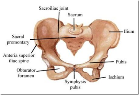
Anatomical position of bony pelvis
- ASIS & Symphysis is on the same coronal plane
- Tip of coccyx & upper margin of symphysis in the same horizontal plane
- So INLET makes an angle of 60 degree with transverse plane
- directed forward
Stages in delivery
- STAGE 1
- cervical dilatation
- starts with onset of true labour pain and ends with full dilatation of the cervix
- STAGE 2
- expulsion of fetus
- Stages
- Descent
- uterine contractions & retractions
- unfolding of fetus
- Engagement
- head engages in the oblique/transverse diameter of the inlet
- suboccopito-bregmatic diameter
- transverse diameter of inlet
- should be more than 9.5cm
- if less cannot accomodate fetal head engagement will fail –> cephalo-pelvic disproportion
- Increased flexion
- Internal rotation
- Extension
- STAGE 3
- expulsion of placenta & membranes
Observe the diameters of the pelvic inlet & outlet. What are the conjugates related to pelvic diameter?
- Anatomical conjugate
- anteroposterior conjugate diameter
- extends from the upper margin of the pubic symphysis to the middle of the sacral promontory
- Obstetrical conjugate
- shortest diameter through which foetal head must pass in it’s course throught the inlet
- measured from middle of back of pubic symphysis to the sacral promontory
- Diagonal conjugate
- anteroposterior diameter of inlet as measured par vaginum
- inability to palpate the sacral promontory suggests that the conjugate diameter of the inlet is adequate for parturition
- palpated means contracted pelvis
- distance between the lower margin of pubic symphysis & sacral promontory
- Subtraction of diagonal conjugate by 1.5cm gives approximate measurement of anatomical conjugate
Pelvis
- Female pelvis inlet: oval
- diameter longer
- transverse widest
- suprapubic angle: 90 degree
- Accomodate gap between thumb & index
- greater sciatic notch: wider
- pelvic cavity: long, conical
- sacrum: short wide
- Male pelvis inlet: heart shaped
- subpubic angle: acute angle
- accomodate gap between index & middle finger
- greater sciatic notch: narrow fish-hook appearance
- pelvic cavity: short, cylindrical
- sacrum: long narrow
Classification of shape of pelvic inlet
- Gynaecoid
- rounded
- Android
- heart shaped (male)
- Anthropoid
- long, narrow, oval
- Platypelloid (flat)
- avoid, long axis tranverse
- like a flat bowl
Supports of the uterus
- Pelvic muscles
- Ligaments
Muscles forming perineal body
Perineal body maintains integrity and should never be torn. If torn, can cause uterus/rectal prolapse. These muscles are supplied by the pudendal nerve.
- levator ani (internal)
- bulbospongiosus (external)
- sphincter ani externus
- transverse perineal
- deep & superficial


Undeniably believe that which you said. Your favorite reason seemed to be at the net the easiest factor to take into accout of. I say to you, I definitely get irked while people consider worries that they just don’t understand about. You managed to hit the nail upon the highest and also outlined out the whole thing with no need side effect , other people could take a signal. Will likely be back to get more. Thank you