Read cases instead!
Disorders of penis
Congenital
- Hypospadias
- ventral (down)
- Epispadias
- dorsal (up)
- Phimosis
- Paraphymosis
Clinical consequences:
Constriction of orifice, urinary tract obstruction/infection, impaired reproductive function
Inflammation
- Balanoposthitis/Balanitis
- inflammation of the glans
- poor local hygience (uncircumcised)
- distal penis swollen (with/without purulent discharge)
- Phimosis
- prepuce cannot be easily retracted over glans
- may be congenital
- associated with balanoposthitis & scarring
- Paraphymosis
- urethral constriction
- Candidiasis
- diabetics
- erosive, painful, pruritic
- can involve entire genitalia
Neoplasia
- Benign
- condylomaacuminatum
- CIS
- Bowen’s disease
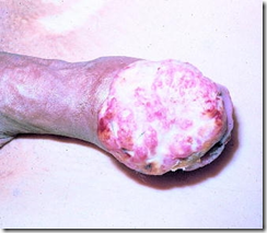
- Squamous cell carcinoma
- uncommon
- uncicumcised men (40-70 yo)
- poor hygiene, smegma, smoking
- human papilloma virus (16 & 18)
- CIS 1st, then progress to SCC
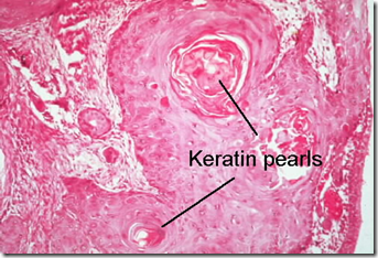
- Microscopic: Keratin pearls
- metastases rare, spread to inguinal lymph nodes
SCC – fungating, exophytic, cauliflower-like
Lesions of scrotum
- tinea crucis/jock itch/inflammation
- superficial dermatophyte infection
- scaly, red, annular plaques, pruritic
- from inguinal crease up to upper thigh
- Peritesticular fibrosis
- scrotal fat necrosis
- fournier’s gangrene
- SCC
- Sir Percoval Pott (Potts cancer)
- Scrotal enlargment
- hydrocele
- accumulation of clear serous fluid within tunica vaginalis
- infections, tumour, idiopathic
- hematocele
- blood in tunica vaginalis
- torsion of testes, direct trauma, hemorrhagic diseases
- chylocele
- filiariasis
- lymph in tunica vaginalis
- differentiate from varicocele
- testicular disease
Disorders of testis
- Congenital
- cryptochidism
- incomplete descent
- 2 phases of descent
- transabdominal
- inguinoscrotal
- fibrosis, firm
- arrest in germ cell development
- marked hyalinzation & thickening of basement membrane of spermatic tubules
- increase in interstitial leydig cells
- greater risk of testicular cancer
- sterility
- absense
- synorchism
- fusion of testes
- cyst
- testicular atrophy
- many causes
- primary testicular atrophy
- due to improper development of testes as a primary failure of genetic origin (kinefelter)
- inflammation
- more common in epididymis than testis
- non specific orchitis
- cystitis
- urethritis
- reach thru vas deferens or lymphatics
- congestion & oedema, infiltration (inflammatory cells)
- fibrous scarring: sterility
- granulomatous (autoimmune) orchitis
- autoimmune
- moderately tender mass
- non tuberculous
- must differentiate from testicular tumour
- affect epididymis 1st:
- gonorrhea
- from posterior urethra
- abscess
- tuberculosis
- caseating granuloma
- affect testis 1st:
- mumps
- post pubertal
- parotid swelling
- sterility
- syphilis
- diffuse interstitial chronic inflammation
- vascular disturbances
Testicular torsion
- Predisposing factors
- incomplete descent
- absence of scrotal ligaments (mobile)
- abnomal attachment of testis to epididymis
- Precipitating cause
- physical trauma
- violent movement
- Pathogenesis
- twisting of spermatic cord
- cut off venous drainage & arterial supply
- vascular engorgement
- venous infarction
- morphology
- intense congestion
- wide spread extravasation of blood into interstitial tissue of testis & epididymis
- hemorrhagic infarction
Testicular tumour
- Germ cells (95%)
- seminoma
- spermatocytic seminoma
- embryonal carcinoma
- AFP +ve, hCG +ve
- yolk sac tumour
- AFP +ve
- choriocarcinoma
- hCG +ve
- teratoma
- more aggressive
- rapid, wide dissemination
- AFP +ve, hCG +ve
- Non germ cell tumour
- leydig cell tumour
- sertoli cell tumour
- benign
- due to steroid use
- endocrinologic syndrome
Clinical view point: seminomatous/ non seminomatous tumours (NSGCT) helps in treatment & prognosis
Clinical features
- painless enlargement of testis
- testicular mass
- biopsy required
- mode of spread
- lymph
- retro peritoneal para-aortic lymph nodes
- spread to mediastinal & supra clavicular nodes
- blood
- lungs (mainly)
- liver
- brain
- bones
Seminoma
- most common germ cell tumour
- adults
- microscopic
- monotonous population of malignant cell
- poorly demarcated lobules
- lymphocytic infiltrate
- gross
- bulky
- grey white
- no haemorrhage/necrosis
- well circumscribed
- homogenous mass
- do not contain alpha-fetoprotein (AFP) or human chorionic gonadotropin (hCG)
Embryonal carcinoma
- numerous necrosis & hemorrhage
- more aggressive than seminoma
- 20-30 yo
- +ve AFP & HCG
- Gross
- smaller in size
- hemorrhagic, necrotic
- poorly demarcated
- microscopic
- cells grow in alveolar/tubular pattern
- papillary convolutions
- primitive grandular differentiation
- undifferentiated
- high mitosis
- tumour giant cells
Yolk sac tumour
- Endodermal sinus tumour
- infants good prognosis
- non encapsulated homogenous yellow white mucinous appearance
- network of cuboidal cells, papillary, solid
- AFP +ve
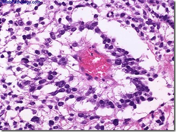
- Schiller Duval bodies
- endodermal sinuses
Schiller Duval body
Choriocarcinoma
- highly malignant
- hCG +ve
- gross
- no testicular enlarment
- small palpable nodule
- hemorrhagic & necrotic
- microscopic
- composed of cytotrophoblasts & synctiotrophoblasts
- mixed pattern
Teratoma
- Contain derivatives of more than 1 germ layer
- endodermal, mesodermal, ectodermal
- common in infants & children
- classified as:
- mature
- common in infants & children
- immature
- with malignant transformations (adults)
- gross
- large
- heterogenous
- solid & cystic areas
- surrounded by well differentiated cartilage & glandular structure
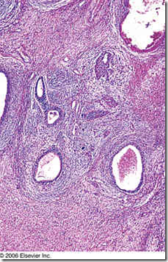
image: mature adult teratoma
image: immature teratoma. hypercellular stroma growing in a concentric fashion around glandular formations
Staging of testicular tumour
- Stage 1
- confined to the testis, epididymis or spermatid cord
- Stage 2
- distant spread
- confined to retroperitoneal nodes
- below the diaphragm
- Stage 3
- metastases outside retroperitoneal nodes
- above the diaphragm
Seminoma tend to be localized to testes for a long time. NSGCT often present with stage 2 & 3. Metastases from seminomas typically involves lymph nodes. Hematogenous spread occurs later in the course of disease. NSGCT metastasize earlier, hematogenous spread earlier, so lungs and liver involved earlier.
Seminomas: extremely radiosensitive. NSGCT: radioresistant
Non-germ cell tumour/sex cord stromal tumours
- Leydig tumour
- granular
- yellowish
- polygonal cells with abundant granular acidophilic cytoplasm
- Sertoli cell tumour
- Gonadoblastoma
Tumour markers
- AFP, HCG, LDH
- seminoma best prognosis
_____________________________________________________________________
EXTRA: LANGE CASE FILES
INTRODUCTION
A 30-year-old man complains of "heaviness" in the scrotal area, which he has noted for about 1 month. He denies any trauma to the area and has no medical problems. He denies the use of tobacco and drinks alcohol occasionally on weekends. On examination, there is a 5-cm firm, nontender area inside the right scrotum. There is no lymphadenopathy.
· What is the most likely diagnosis?
· What is the most likely histologic finding?
ANSWERS TO CASE 11: Testicular Cancer
Summary: A 30-year-old male male has a 1-month history of right scrotal "heaviness." Examination reveals a 5-cm firm, nontender area inside the right scrotum. There is no adenopathy.
· Most likely diagnosis: Testicular cancer
· Most likely histologic finding: One or more germ cell tumor types, including seminoma, embryonal carcinoma, choriocarcinoma, yolk sac tumor, and teratoma.
CLINICAL CORRELATION
Introduction
This patient, a young man with "scrotal heaviness," represents a very typical presentation of a testicular mass. Not all masses in the scrotum are testicular in nature. Physical examination and ultrasound of the scrotum often help differentiate a solid testicular mass from benign entities such as hydrocele (water sac around the testicle), varicocele (dilated testicular veins), and epididymoorchitis (infection and/or inflammation of the epididymis and testis).
A hard painless mass within the testicle should be considered testicular cancer until proved otherwise. Such a mass often is overlooked by the patient until it is discovered accidentally or brought to his attention by an unrelated regional occurrence such as minor trauma to the scrotum. Germ cell tumors of the testis peak in men between ages 15 and 40 years. Seminoma, embyronal carcinoma, choriocarcinoma, yolk sac tumor, and teratoma can appear in a pure or, more often, mixed form. Characterizing the tumor awaits review of the surgical specimen and is not possible before orchiectomy (surgical removal of the testicle). Although cure of testicular tumors now occurs in the high 90 percent range, because the malignancy affects primarily young men, issues of compromised fertility, complications of therapy, and surveillance for recurrence and second malignancies are matters of concern.
Approach to Testicular Tumors
Definitions
Cryptorchidism: Congenital failure of one or both testes to descend from the abdominal (in utero) position to the scrotal postnatal position. Cryptorchid testicles have a higher incidence of malignancy, which is not reduced by surgical relocation into the scrotum. Patients who have had surgical correction of cryptochid testes require intensive education about the importance of self-examination because of the increased risk of malignancy.
Radical orchiectomy:Removal of the testicle, epididymis, and testicular cord up to the internal inguinal ring. This operation is performed through an incision in the inguinal area that is similar to the incision made to repair inguinal hernias.
Nonseminomatous germ cell tumors: Because of the distinct difference in treatment of a pure seminoma
, germ cell tumors often are divided into two categories: (1) pure seminomas and (2) nonseminomatous germ cell tumors (all other pure tumors or seminomas with a mixed component). Pure seminomas are very radiosensitive, whereas nonseminomatous tumors are radioresistant (see Table 11-1 for classification).
Tumor markers: Substances secreted by cancers that can be detected and measured to assess the presence and extent of the primary, metastatic, or recurrent disease. Alpha-fetoprotein and human chorionic gonadotropin beta are tumor markers for testicular cancer.
Retroperitoneal lymph node dissection (RPLND): Removal of lymph nodes that provide the primary lymphatic drainage from the testes from the bifurcation of the great vessels up to and beyond the renal hilum. This procedure may be curative in cases of low-volume metastatic disease, because most germ cell tumors progress in an orderly fashion from primary intratesticular to lymphatic before metastasis to solid organs.
Discussion
Any male presenting with an intrascrotal mass requires a thorough physical examination of the genitalia. The examination should be performed with the male standing as well as lying flat. The primary goal is to determine whether the mass lies within the testicle or is extratesticular. Masses within the testes require further evaluation. Scrotal ultrasound is an excellent study to confirm the presence of and characterize a suspected testicular mass. If an intratesticular mass is confirmed on examination and/or ultrasound study, the diagnosis of a germ cell tumor is made by inguinal orchiectomy and pathologic analysis of the testis. Once the diagnosis of a germ cell tumor is confirmed and characterized by the histology, tumor staging is performed.
Before an inguinal orchiectomy, serum markers are drawn, such as alpha-fetoprotein and human chorionic gonadotropin beta levels, if a germ cell tumor is confirmed. A CT scan of the abdomen is obtained after the inguinal orchiectomy, looking for the presence of enlarged lymph nodes in the retroperitoneum. The use of intravascular contrast material helps outline the aorta and vena cava, on which lies the primary lymphatic drainage echelon of the testis. A chest radiograph is obtained with additional views if needed. These staging studies augment the physical examination, which assesses for abdominal masses, breast enlargement (caused by the influence of serum tumor markers), and the Virchow lymph node in the supraclavicular region.
Patients with pure seminoma and disease that is thought not to be metastatic are treated with a short course of radiotherapy to the retroperitoneal and ipsilateral iliac lymph nodes. Such simple therapy yields 90 percent 5-year disease-free survival. For patients with mixed germ cell tumors, treatment depends on the presence and extent of metastatic disease. Retroperitoneal lymph node dissection may be chosen as a therapy when there is no evidence of gross metastatic involvement. Surgery will define the extent of disease and in some cases may cure the malignancy.
For patients for whom chemotherapy is chosen, a variety of chemotherapeutic protocols exists that are administered using x-ray and serum studies to monitor effectiveness. Sometimes metastatic deposits will be reduced but not eliminated by the chemotherapy, requiring surgical excision.
The five types of germ cell tumors: seminoma, embryonal carcinoma, choriocarcinoma, yolk sac tumor, and teratoma are collectively responsible for about 90 percent of testicular tumors. The remaining testicular tumors originate from the gonadal stroma, such as Leydig and Sertoli cells. A variety of rare tumors occur with a frequency far less than that of the more common germ cell tumors. The histologic subtype of germ cell tumors is an important distinction, because therapy is predicated on a complete examination of the entire tumor to determine whether it is of pure type or mixed in origin. Pure seminomas are distinct from other solid tumors of the testicle in that seminomas are exquisitely radiosensitive. Other germ cell tumors, whether in pure or mixed form, do not have sufficient radiosensitivity to make radiation therapy a commonly employed treatment modality.
Whatever combination of radiation, surgery, and/or chemotherapy is used, patients with testis tumor often are cured of their malignancies. After a diagnosis of testis cancer, the patient is warned that therapies, especially chemotherapy, can have a negative long-term impact on fertility. Some patients choose to bank sperm before the initiation of chemotherapy. All patients with testis tumor have at least a 5-year period of intensive monitoring on or off therapy. Compliance with the rigorous program of physical examinations, x-rays, and blood tests must be stressed to the patient. Radiation, surgery, and chemotherapy have their own distinct set of side effects, which, depending on the patient’s age, may be not only short-term but also long-term, revealing themselves many decades later.
COMPREHENSION QUESTIONS
[11.1] A 35-year-old man presents with the painless enlargement of one testicle. Physical examination finds a single nontender testicular mass that measures about 3 cm in the greatest dimension and does not transilluminate. What type of testicular tumor is most likely to be present in this individual?
A. Germ cell tumor
B. Gonadal stromal tumor
C. Mesenchymal tumor
D. Sex cord tumor
E. Surface epithelial tumor
[11.2] What type of primary testicular tumor is most radiosensitive?
A. Seminoma
B. Embryonal carcinoma
C. Yolk sac tumor
D. Choriocarcinoma
E. Immature teratoma
[11.3] A 27-year-old man has surgery for a testicular mass. Histologic sections reveal the mass to be a testicular yolk sac tumor. Which one of the substances listed below is most likely to be increased in this patient’s serum?
A. Acid phosphatase
B. Alkaline phosphatase
C. Alpha-fetoprotein
D. Human chorionic gonadotropin
E. Prostate-specific antigen
ANSWERS
[11.1] A. An intratesticular mass in an adult, especially a mass that does not transilluminate, should be considered a testicular tumor until proved otherwise. The two main types of primary testicular tumors are germ cell tumors and sex cord gonadal stroma. Germ cell tumors are more common and have a peak incidence in men between 15 and 40 years of age.
[11.2] A. The histologic classification of testicular germ cell tumors includes seminomas, embryonal carcinomas, yolk sac tumors, choriocarcinomas, and teratomas. It is important, however, to group these testicular tumors into two general categories: those which are pure seminomas and those tumors which contain nonseminomatous elements. The reason for this classification is the fact that pure seminomas are very radiosensitive and nonseminomatous tumors are radioresistant and require the use of chemotherapy.
[11.3] C. Tumors sometimes secrete specific substances that appear in the serum. These tumor markers can be used clinically for diagnosis, for staging, and for following patients after surgery to look for recurrences of the tumor. Two important tumor markers associated with testicular neoplasms are alpha-fetoprotein (AFP) and human chorionic gonadotropin beta (hCG-beta). Increased levels of AFP are associated with embryonal cell carcinoma or endodermal sinus tumor, whereas increased levels of hCG are associated with choriocarcinoma. Increased levels of both AFP and hCG are seen with mixed germ cell tumors. It is important to not
e that although seminomas may be associated with increased levels of hCG, pure seminomas are never associated with increased levels of AFP.
PATHOLOGY PEARLS
· The majority of testicular tumors are germ cell tumors.
· Seminomas are very radiosensitive.
· An intratesticular mass should be considered a testicular tumor until proved otherwise.


