Read written notes.
Some images on cerebellum:


Location of cerebellum:
- Largest part of hindbrain
- Occupies most of posterior cranial fossa
- Lies behind pons & medulla
- forming roof of 4th ventricle
- Separated from posterior part of cerebrum
- by tentorium cerebelli
_____________________________________________________________________
Cerebral peduncles:

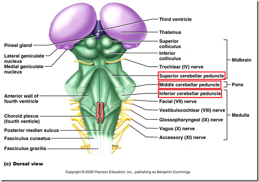
Joined to the brain stem via:
- Superior cerebellar peduncle –> Midbrain
- Middle cerebellar peduncle –> Pons
- Inferior cerebellar peduncle –> Medulla
Fibres in peduncles:
- Superior peduncle
- ventral spino-cerebellar
- only spinocerebellar tracts go thru sup peduncle
- cerebello-thalamic
- cerebello-rubral (red nucleus)
- ventral spino-cerebellar
- Middle peduncle
- ponto-cerebellar
- Inferior peduncle
- vestibulo-cerebellar
- vesticular nuclei
- posterior spino-cerebellar
- lower limb
- cuneo-cerebellar
- upper limb
- reticulo-cerebellar
- reticular nuclei
- olivo-cerebellar
- olive nuclei
- vestibulo-cerebellar
_____________________________________________________________________
Gross Anatomy
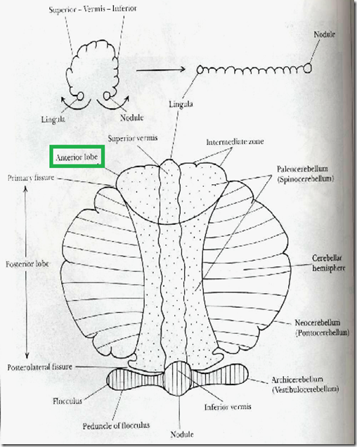
- Shape: Ovoid
- 2 cerebellar hemispheres
- united by a constricted median part
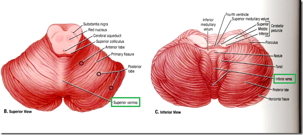
- Vermis (spine of cerebellum)
- Superior vermis
- Inferior vermis
- Vermis (spine of cerebellum)
- united by a constricted median part
- Inferior aspect
- 2 halves are separated by a hollow: VALLECULA
- Hemispheres show parallel fissures
- separating thin rounded FOLIA
- similar to cerebral sulci/gyri
Lobes & Fissures
- Hemispheres divided into
- 3 lobes
- Middle lobe: largest part
- 2 fissures
- Primary Fissure
- V-shaped with apex directed backwards
- Separates anterior lobe from middle lobe
- Horizontal fissure
- deep along posterior margin
- separates superior & inferior surface
- no functional significance
- Primary Fissure
- 3 lobes
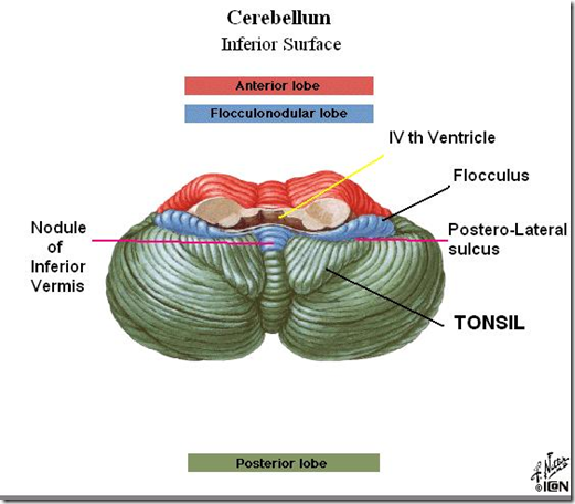
- Posterolateral fissure
- separates middle lobe from floncculonodular lobe
- Flocculonodular lobe (hidden in the inferior part)
- laterally placed small ovoid flocculus connected medially to nodule (on inferior vermis)
- Neocerebellum: helps cerebral cortex in timing & program complex activities
_____________________________________________________________________
Phylogenetic & Functional Parts

- Flocculonodular lobe
- smallest
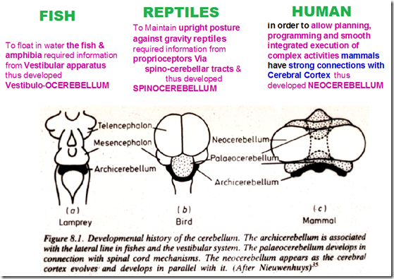
- constitute phylogenetic archicerebellum
- functional vestibulo-cerebellum
- primitive, old
- still reacting to stimuli from balance organ vestibule & 8th nerve
- Anterior lobe
- antero-superior part
- includes most of superior vermis & anterior-most part of superior surface
- Anterior lobe + (Pyramid + Uvula of vermia)
- constitutes phylogenetic paleocerebellum
- functional spino-cerebellum
- reacting to simuli from proprioceptive endings in muscles and tendons
- spino-cerebellar tracts, reticulo-cerebellar tracts
- constitutes phylogenetic paleocerebellum
- Middle lobe (also known as posterior lobe)
- a large part of cerebellar hemisphere
- entire inferior surface & most of superior surface
- behind primary fissure
- constitute phylogenetic neocerebellum
- functional cerebro-cerebellum
- reacting to stimuli from opposite cerebral cortex about planned movements
- eg. right cerebral cortex control kicking of left leg.
- if not developed properly, difficulty in kicking left leg. vice versa
- a large part of cerebellar hemisphere
_____________________________________________________________________
Functional Anatomy & Afferent, Efferent Connections of Cerebellum
Vestibulocerebellum (Flocculonodular lobe)
- responds to vestibular stimuli from internal ear
- assists in maintaining equilibrium by modification in muscle tone
- postural muscles
- eye muscles
- trapezius
- sternomastoid
- erector spinae
- Main deciding factor: fastigial nucleus
- which deep nucleus controls equilibrium: fastigial nucles
- Afferent:-
- Vestibulo-cerebellar
- via inferior cerebellar peduncle
- from vestibular nuclei in the floor of 4th ventricle
- also from vestibular nerve (8th nerve)
- Vestibulo-cerebellar
- Efferent:-
- Cerebello-vestibular
- via inferior cerebellar peduncle
- mainly to lateral vesticular nucleus
- Cerebello-reticular
- reticular nucleus brain stem
- ——> then via vestibulo-spinal & reticulo-spinal tract modify the lower motor neurons
- Cerebello-vestibular
Spinocerebellum (Anterior lobe)
Paleocerebellum
- Responds to
- proprioception input from muscle spindle
- touch/pressure input
- Maintains posture of the body by
- modifying muscle tone
- regulate voluntary movements
- Afferent:-
- from spinal cord
- from lower limb, trunk
- via posterior spino-cerebellar tract
- from upper limb, thorax
- via anterior spino-cerebellar tract
- via cuneo-cerebellar tract
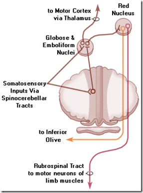
- Efferent:-
- cerebello-cerebral
- via thalamus
- cerebello-reticular
- cerebello-rubral
- to red nucleus in midbrain
- ——–> then via reticulo-spinal & rubro-spinal tract modify the gamma motor neurons in spinal cord
- modify muscle tone
- cerebello-cerebral
Neocerebellum
- receives input from cerebral cortex & olivary nucleus
- facilitates smooth coordinated voluntary movements
- ensures that the force, direction & extent are accurate
- role in motor learning (learn diff movements)
- athletes
- timing & amplitude of learned movements are encoded here
- diverse cognitive functions
- attention
- processing of language
- any smooth movements require perfect synergism between agonist & antagonist group of muscles
- Afferent:-
- Cortico-ponto-cerebellar
- via middle cerebellar peduncle
- from motor areas of opposite cerebral cortex after synapse in pontine nuclei
- there fibres – anatomical basis of control of cerebellar activity by cerebral cortex
- Olivo-cerebellar
- via inferior cerebellar peduncle
- from inferior olivary nucleus in medulla
- Cortico-ponto-cerebellar
- Efferent:-
- Cerebello-thalamic
- via superior cerebellar peduncle
- the to motor cortex
- via cortico-spinal tract & cortico-nuclear fibres
- coordinate voluntary movements
- Cerebello-rubral
- via superior peduncle
- to red nucleus
- Cerebello-thalamic
_____________________________________________________________________
Histology
- Similar to cerebral cortex
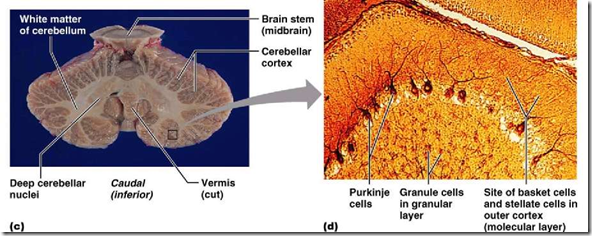
- central white matter
- peripheral grey matter
- Midsagittal section
- multiple folia containing core of white matter covered superficially by grey matter
- Central core of white matter contains
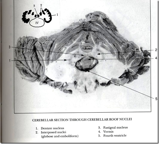
- 4 intra-cerebellar nuclei
- receiving input from cerebellar cortex
- extending outflow (axons) via superior & inferior peduncles
- 4 intra-cerebellar nuclei
MEDIAL TO LATERAL
- Fastigial nuclei (medial)
- primitive
- receives input from vermis (archicerebellum)
- Globose, emboliform nuclei
- together known as interpositus
- receive input from paravermal area (spinocerebellum)
- Dentate nuclei (lateral)
- receives input from most of cerebellar hemisphere (neocerebellum)
- largest nucleus
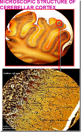
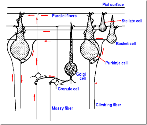
Microscopic structure of cerebellar cortex (3 layers)
- External Molecular layer
- Middle Purkinje layer
- large flask shaped neurons
- arranged in single tier
- in a plane transverse to folium
- dendrites produce profuse branching
- receive communications afferent fibres entering cerebellum
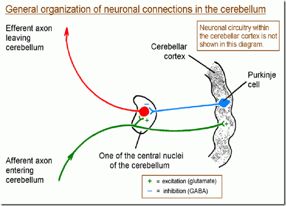
- axons run to DEEP CEREBELLAR NUCLEI
- sole output of cerebellar cortex
- functionally
- inh
ibitory (GABA)- stimulated purkinje inhibits deep nuclei
- inh
- Internal Granular layer
- closely packed small neurons
All afferent input via 2 different fibres:
- Mossy fibre
- entire inflow of cerebellum
- except from inferior olive
- particularly from cerebral cortex via pons
- they synapse in expanded MOSS-like appearance with granule & golgi cells in granular layer
- fast-firing
- rapid adjustment for ongoing movement
- entire inflow of cerebellum
- Climbing fibre
- inflow from inferior olive
- run up and synapse with dendrites of single Purkinje cell
- slow-firing
- helps in learning muscle habits
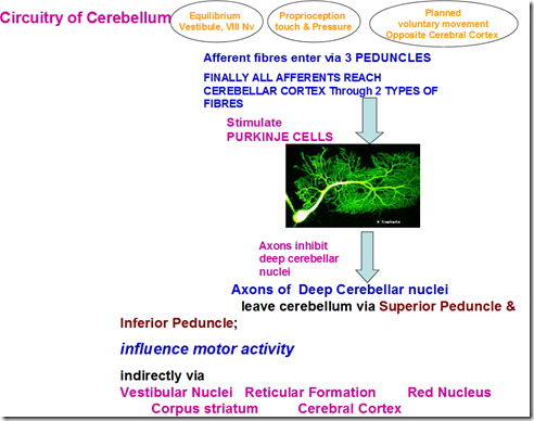
Circuitry of Cerebellum
Process:
- Afferent
- equilibrium, vestibule, 8th nerve
- proprioception, touch, pressure
- planned voluntary movement, opposite cerebral cortex
- enter via 3 peduncles
- all afferents reach cerebellar cortex through 2 types of fibres
- mossy fibre
- climbing fibre
- stimulate purkinje cells
- axons inhibit cerebellar nuclei
- leave cerebellum via superior peduncle & inferior peduncle
Influence motor activity, indirectly via:
- vestibular nuclei
- corpus nuclei
- red nucleus
- reticular formation
- cerebral cortex
_____________________________________________________________________
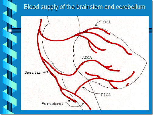
Blood supply
- Superior surface
- Superior cerebellar branch of basilar artery
- Inferior surface (Anterior part)
- Anterior inferior cerebellar branch of basilar artery
- Inferior surface (Posterior part)
- Posterior inferior cerebellar branch of verterbral artery
Veins
- Straight sinus
- Occipital sinus
- Transverse sinus
*Trombosis will affect many organs – because brainstem controls many important activities of the body
_____________________________________________________________________
Vestibulo-Cochlear apparatus
- Lateral line of organ of fish and amphibian
- enable fish to keep upright
Human vestibular apparatus
- vestibule, vestibular nerve, vestibular nucleus, cerebellum, vestibulospinal tract, superior colliculus, medial longitudinal fasciculus, tectospinal tract, reticulospinal tract
- responsible for maintanence of equilibrium
- based on perceived movements of the head
- Reflex adjustments
- muscle tone
- muscle tension
- muscle contraction
- Muscles involved
- neck muscles
- movement of head
- muscle of spine
- resist pull of gravity
- extraocular muscle
- maintain movement of eyes with head position
- neck muscles
_____________________________________________________________________
Vestibular disorders
Disturbance of vestibular function:
- Central
- brain stem vestibular nuclei affected
- Peripheral
- labyrinth (internal ear)
- vestibular component of 8th nerve
Giddiness (Vertigo)
- Nystagmus (check vestibular function)
- disturbance of reflex control of extraocular muscles
- vestibulo-ocular reflex
- involuntary to-and-fro movement (rhythmic) of the eyes
- Types:
- Vestibular nystagmus
- uncontrollable rhythmic oscillation of the eyes
- fast phase is away from the side of lesion
- central lesion: spontaneous nystagmus without a cognitive, visual or vestibular stimulus
- see nystagmus even without stimulus
- Cerebellar nystagmus
- disorder of cerebellum by tumour, thrombosis, ischaemia, trauma)
- unilateral lesion
- amplitube greater when eyes rotated to the side of lesion
- can be horizontal/vertical
- Vestibular nystagmus
- disturbance of reflex control of extraocular muscles
- Ataxia
- incoordination / clumsiness of movements
Signs of cerebellar disorder
- Always ipsilateral (same sided)
- No paralysis
- No sensory loss
- Wide-based gait
- on walking, staggers to the affected side
- Hypotonia
- shaking limb, excessive movement of terminal joints
- Ataxia
- disturbance of coordinated voluntary movement
- tremor on fine movements
- writing
- shaving
- buttoning
- Finger-nose test
- past-pointing to ear instead
- Heel-knee test
- unable to do properly
- Pendular knee reflex
- continue longer
- Dysdiadochokineses
- on pronation & suppination of forearm rapidly
- imcomplete & jerky movement




thanks for taht nice overvview, good diagrams as well.
Pete D
pretty okay