
- OLIS
- Composition of cartilage
- Joints
- Dr Nilesh’s blog – knee joint
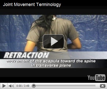
Before you start learning about the joints itself, it is important to learn about the joints articulation/movements so that you will understand better how the different types of joints result in those movements.
- Extension & Flexion
- Abduction & Adduction
- Circumduction
- all of the above
- Medial & Lateral rotation
- Lateral flexion
- Pronation & Supination
- Eversion & Inversion
- Protraction & Retraction
- Elevation & Depression
- Opposition
- Dorsiflexion & Plantarflexion
_____________________________________________________________________
Joints
Definition: Is the site where 2 bones meet.

Classification of joints
- Functional classification (based on degree of movement)
- Synarthroses
- no movements at all
- Amphiarthroses
- Slight movements
- Diarthroses
- Freely movable
- Synarthroses
- Structural classification
- Fibrous joints
- bones joined by fibrous tissue
- Categories
- Sutures
- skull bones
- Syndesmoses
- inferior tibio-fibular joint
- Gomphoses
- tooth sockets
- Sutures
- Cartilaginous joints
- bones joined by cartilage
- Categories
- Synchondroses (Primary)
- No movements
- Epiphyseal plate (hyalin cartilage)
- Symphyses (Secondary)
- Slight movements
- Intervertebral discs
- Synchondroses (Primary)
- Synovial joint
- bones separated by fluid containing cavity
- Examples:
- Knee
- Elbow
- Contains
- Articular cartilage
- hyaline cartilage
- covers the bone surfaces
- Joint cavity
- Articular capsule
- Fibrous capsule
- Synovial membrane
- Synovial fluid
- Reinforcing ligaments
- Articular cartilage
- Types of synovial joint
- Plane joint
- intercarpal joint
- gliding movement
- nonaxial
- Hinge joint
- elbow joint
- extend and flex only
- uniaxial
- Pivot joint
- proximal radio-ulnar joint
- rotation
- uniaxial
- Condyloid joint
- metacarpo-phalangeal joint
- biaxial
- Saddle joint
- carpo-metacarpal joint
- only in thumb
- biaxial
- Ball & Socket joint
- shoulder joint
- all movements
- multiaxial
- Plane joint
- Fibrous joints

Axis of joint movements
- Non-axial
- only slipping possible
- Uniaxial
- movements in 1 plane
- Biaxial
- movements in 2 planes
- Multiaxial
- movements around all 3 plants
Reinforcing ligaments
- Capsular
- thickening of the capsule
- hip joint
- Extracapsular
- shoulder joint
- Intracapsular
- knee joint
Bursae & Tendon sheaths
- Bursae
- flattened fibrous sac lined with synovial membrane and filled with synovial fluid
- Tendon sheath
- elongated bursae into which a tendon sheath is invaginated
Purpose: Reduce friction between the bones.
Stability of joints
If stability is not maintained, joint can get dislocated.
- Articular surfaces
- if articular surfaces are fitting together, stability is increased
- Ligaments
- surr
ounding the joints provides stability - the greater the number of ligaments, the stronger they are
- more stable
- surr
- Surrounding muscles and their tone
- tendon and muscles crossing the joints stabilize the joint
Blood supply of joints
- From the arteries surrounding the joint
- articular arteries
- forms anastomosing network
- articular arteries
Nerve supply of joints
- From the nerves supplying the muscles which crosses the joints
- Hilton’s law
_____________________________________________________________
Shoulder Joint (Glenohumeral joint)


Characteristics
- Synovial joint
- Ball & Socket joint
- Multiaxial
- Ball: Humerus
- Socket: Glenoid cavity
- pear shaped
- shallow
- deepened by a rim of fibrocartilage attached to the rim of glenoid cavity
- glenoidal lambrum
- deepened by a rim of fibrocartilage attached to the rim of glenoid cavity
- not reciprocal
- only in contact with 1/3 of humeral head
Stability of joint

- Muscle: Rotator cuff (of Codmann)
- important due to ligament laxity
- All sides except inferior part (SITS)
- From Infront
- Subscapularis
- From behind
- Infraspinatus
- Teres Minor
- From above
- Supraspinatus
- From Infront
- Ligaments
- Capsular ligament (Glenohumeral ligament)
- Superior
- Middle
- Inferior
- Generally lax/loose
- Tightens only at extreme of movements
- Capsular ligament (Glenohumeral ligament)


Reasons for shoulder instability:
- *No reciprocal curvature of articular surfaces
- Ligaments weak and lax
- Gravity
Factors maintaining shoulder stability:
- Muscles and Tendons
- Yes
- Surface tension & vacuum
- Synovial fluid
- Adhesive & Cohesive Force
Bones- Almost nil
Ligaments- Negligible
Shoulder joint movements & muscle involvement
- Transverse axis
- Flexion
- Deltoid (anterior fibres)
- Pectoralis major (clavicular head)
- Extension
- Deltoid (posterior fibres)
- Latissimus dorsi
- Flexion
- Anteroposterior axis
- Adduction
- Pectoralis major
- Latissimus dorsi
- Teres major
- Triceps brachii (long head)
- Abduction
- Deltoid
- Supraspinatus
- Serratus anterior
- Trapezius (upper and lower fibres)
- Adduction
- Vertical axis
- Medial rotation
- Pectoralis major
- Deltoid (anterior fibres)
- Latissimus dorsi
- Teres major
- Lateral rotation
- Deltoid (posterior fibres)
- Infraspinatus
- Teres minor
- Medial rotation
- Circumduction
- occurs at 180 degrees
- movement partly at shoulder joint, partly at pectoral girdle
- First 15 – 30 degrees
- Supraspinatus
- Up til 90 degrees
- Deltoid
- 90 – 180 degrees
- Roration of scapula
- Trapezius
- Serratus anterior
- Roration of scapula
- occurs at 180 degrees
_____________________________________________________________________
Hip Joints (Acetabulofemoral joint)

Characteristics
- Synovial joint
- Multiaxial
- Ball & Socket joint
- Ball: Femoral head
- covered by hyalin cartilage except for fovea
- Socket: Acetabulum
- cup shaped
- large & deep
- narrow mouth
- mouth made narrower by a rim of fibrocartilage which is attached to the rim of acetabular cavity
- acetabular labrum
- mouth made narrower by a rim of fibrocartilage which is attached to the rim of acetabular cavity
- articular surface
- horse-shoe shaped
- Reciprocally curved
- Ball: Femoral head
Hip joint stability


- Muscles
- Lateral
- Tensor fasciae latae muscle
- Iliotibial tract
- Vastus lateralis muscle
- Posterior
- Gluteus maximus
- Hamstring muscle
- Semitendinous muscle
- Semimembranous muscle
- Biceps femoris muscle
- Adductor magnus muscle
- Gracilis muscle
- Deep
- Gluteus medius
- Obturator gamellus muscle
- Obturator internus muscle
- Inferior gamellus muscle
- Quadratus femoris muscle
- Biceps femoris muscle (long head)
- Semitendinous muscle
- Semimembranous muscle
- Lateral
- Ligaments
- Iliofemoral
- Ischiofemoral
- Pubofemoral
- Ligamentum teres
- ligament of head of femur
- Transverse acetabular


Hip joint movements
- Same as movements of shoulder joints
Factors for hip joint stability
- Shape
- Reciprocal curvatures
- The articular surfaces are configured and positioned in such a way that normal loading enhances the closeness of their fit
- better than shoulder joint
- Reciprocal curvatures
- Ligaments
- major stability influence
- guide and align normal joints through their range of motion
- strong & inelastic structure
- limit articular motion
- iliofemoral joint one of the strongest in the body
- thick capsule
- major stability influence
- Muscles
- yes, but not primary form of support
- primary role
- steady the pelvis on the lower limbs
- maintain posture
- Surface tension
- Body weight
_____________________________________________________________________
Knee joint
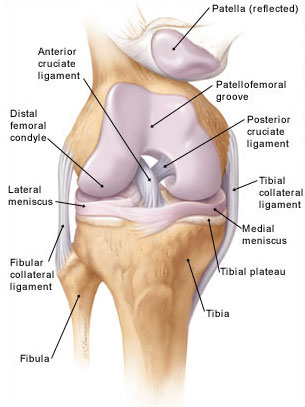

Characteristics
- Largest joint in the body!
- Synovial joint
- Modified hinge joint
- Composed of 3 joints
- Condylar
- Medial femorotibial
- Lateral femorotibial
- Saddle
- Femoropatellar
- Condylar
- Articular surfaces
- Condyles of femur
- Condyles of tibia
- Patella
- Mobility
- by bones
- Stability
- by soft tissues
Stability of joint

- Ligaments
- Fibrous capsule
- Only in posterior
- deficient in front
- Pierced by
- suprapatellar bursae
- tendon of popliteus
- Only in posterior
- Patellar Ligament / Ligamentum patellae
- Tendon of quadratum femoris
- Tibia collateral ligament
- From medial epicondyle of femur
- To tibia
- Fibular collateral ligament
- From lateral epicondyle of femur
- To head of fibula
- Anterior cruciate ligament
- Named by attachment to tibia
- Taut in extension
- Posterior cruciate ligament
- Named by attachment to tibia
- Taut in flexion
- Coronary ligament
- Oblique popliteal ligament
- Arcuate popliteal ligament
- Fibrous capsule
- Muscles
- Extensors
- Quadriceps femoris
- Tensor fasciae latae
- Flexors
- Hamstrings
- Biceps
- Semitendinosus
- Semimembranosus
- Hamstrings
- Medial Rotators
- Popliteus
- Semitendinosus
- Semimembranosus
- Lateral Rotators
- Biceps femoris
- Extensors
Movements of muscles
- Extension, Flexion
- Medial & Lateral rotators
- Screw home movement
- locking/unlocking
- This is the name given to the action when a knee reaches full extension and just as the knee locks into hyperextension the knee rotates in an outward direction just a few degrees. This has the effect of putting most of the weight onto the cartilage, menisci, and bones of the joint while giving a rest to the muscles of the thigh and ca
lf. What allows for this is that the medial femoral condyle is slightly curved and the medial condylar notch is slightly longer than the lateral condylar notch
- Accessory movements
- only on partially flexed knee
- Wider range of rotation
- AP gliding of tibia on femur
- Some amount of abduction & adduction
- Some amount of separation of tibia from femur
Factors maintaining stability of knee joint
- Anteroposterior stability
- Cruciate ligaments
- Side-to-side stability
- Collateral ligaments
- Capsular ligaments
- weak
- reinforced by tendinous expansions
- During partial flexion
- Iliotibial tract
Reasons for instability of knee joint
- Due to incongruence
- Tibial condyles too small & shallow for the large & convex femoral condyles
- Angulation of thigh on leg
- shallow surfaces of patellar & femur
Menisci


- Menisci are semilunar fibrocartilages
- that try to deepen the articular surfaces of the tibial condyles
- 2 attached ends
- 2 borders
- 1 fixed
- 1 free
- 2 surfaces
- 1 concave
- 1 flat
- 2 parts
- outer vascular
- inner avascular
- Medial meniscus
- Semicircular
- attached to the tibial collateral ligament
- Lateral meniscus
- Circular
- attached to medial condyle of femur by
- 2 meniscofemoral ligaments
- attached to popliteal tendon (tendon of popliteus)
Clinical anatomy (look up later)

- Q angle
- Genu valgum
- angle less than 165 degrees
- Genu varum
- angle more than 180 degrees
- Genu valgum
- Knee jerk
- Osteoarthritis
- Meniscal tears
- Cruciate ligament injury
- Bursitis
- Patellar fracture
- Xray


_____________________________________________________________________
Ankle joint (Talocrural joint)
 Characteristics
- Synovial joint
- Hinge joint
- Made up of 3 bones
- Tibia
- Fibular
- Talus
- distributes the weight
Stability
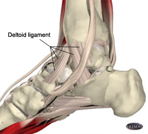
- Strong structure
- Strong ligaments
- Medial collateral ligament (Deltoid ligament)
- tibionavicular ligament
- calcaneotibial ligament
- anterior talotibial ligament
- posterior talotibial ligament
- Lateral collateral ligament
- anterior talofibular ligament
- calcaneofibular ligament
- talocalcaneal ligament
- posterior talocalcaneal ligament
- posterior talofibular ligament
- Medial collateral ligament (Deltoid ligament)
- Tendinous reinforcement
Movements (2 only)
- Dorsiflexion
- Plantarflexion
_____________________________________________________________________
Joints of the foot

- Tibiofibular joints
- Superior
- Middle
- Inferior
- Intertarsal
- Subtalar joint (Talocalcanean)
- Talocalcaneonavicular joint
- Calcaneocuboid joint
- Transverse tarsal joint
- Smaller joints
- Cuneonavicular
- Cuboideonavicular
- Intercuneiform
- Cuneocuboid
- Tarsometatarsal
- Intermetatarsal
- Metatarsophalangeal
- Interphalangeal
Movements of the foot
- Inversion (plantarflexion + adduction of foot)
- Eversion (plantarflexion + abduction of foot)
- Subtalar joint
- Talocalcaneonavicular joint
- calcaneum and navicular move medially/laterally around the talus
- carrying the foot with them
- calcaneum and navicular move medially/laterally around the talus
- Metatarsophalangeal joints (ellipsoidal joints) – movements
- Extension
- Flexion
- Abduction
- Adduction
- Interphalangeal joints (hinge joints)
- Extension
- Flexion
Arches of the foot

- Longitudinal arch
- Medial longitudinal arch
- Posterior pillar
- Calcaneum
- Anterior pillar
- Navicular
- Cuneiforms
- Medial 3 metatarsals
- Summit
- Talus
- Posterior pillar
- Lateral longitudinal arch
- Posterior pillar
- Calcaneum
- Anterior pillar
- Cuboid
- Lateral 2 metatarsals
- Summit
- Calcaneo-cuboid articulation
- Posterior pillar
- Medial longitudinal arch
- Transverse arch
Clinical anatomy of the arches
- Footprint
- Pes planus
- flat foot
- Pes cavus
- high arch of the foot that does not flatten with weight bearing
- Talipes equinovarus
- club foot
_____________________________________________________________________
Elbow joint

Characteristics
- Synovial joint
- Hinge joint
- Composed of 2 joints
- radio-capitulum joint
- articular surfaces:
- head of radius
- capitulum (of humerus)
- articular surfaces:
- humero-ulnar joint
- articular surfaces:
- Trochlea notch of ulna
- Trochlea of humerus
- articular surfaces:
- radio-capitulum joint
- Elbow joint continuous with radio-ulnar joint
- Bony landmarks
- Humerus
- Radial fossa
- Coronoid fossa
- Olecranon fossa
- Humerus

Stability

- Ligaments
- Capsule
- Superiorly
- encloses capitulum, trochlea, radial fossa, coronoid fossa & olecranon fossa
- Inferiorly
- attached to the trochlea notch & annular ligament
- Superiorly
- Radial collateral ligament
- fan shaped
- attached to
- lateral epicondyle
- annular ligament
- Ulnar collateral ligament
- Triangular
- anterior, posterior & oblique bands
- attached to
- medial epicondyle
- ulna
- Triangular
- Capsule
Blood & Nerve supply of the elbow joints
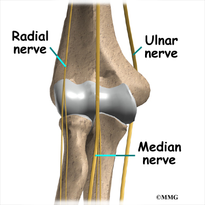
- Blood supply
- anastomosis around the joint
- Main artery
- brachial artery
- branches into collateral arteries
- Nerve supply
- Ulnar nerve
- Median nerve
- Radial nerve
- Musculocutaneous nerve
Movements of elbow joints
- Flexion
- Brachialis
- Biceps
- Brachioradialis
- Extension
- Triceps
- Anconeus
Relations
- Biceps tendon
- Brachial artery
- Median nerve
Elbow joint – Carrying angle

- Transverse axis of joint directed medially and laterally
- Due to this,extended forearm not in line with arm
- makes an angle of about 163 degrees
- Disappears in
- full flexion
- pronation
- Anomaly
Clinical anatomy
- Distension
- occurs posteriorly due to effusion
- aspirated posteriorly
- Dislocation
- usually posteriorly
- associated with fracture of coronoid
- triangular relationship is lost between
- Olecranon
- 2 epicondyles
- Subluxation of head of radius
- occurs in children
- when arm is pulled in pronation
- radial head slips out from annular ligament
- Tennis elbow
- abrupt pronation
- results in tear of radial collateral ligament
- or extensor ligaments originating from lateral epicondyle
- abrupt pronation
_____________________________________________________________________
Radio-ulnar joint

Characteristics
3 joints together
- Superior
- Synovial joint
- pivot type
- Articular surfaces
- circumferance of the head of radius
- osseofibrous ring
- formed by radial notch of ulnar & annular ligament
- Blood supply
- anastomosis around the lateral side of elbow joint
- Nerve supply
- Median nerve
- Radial nerve
- Musculocutaneous nerve
- Movements
- Suppination
- Palms facing upwards
- Antigravity (more powerful)
- Muscles
- Biceps muscle
- Suppinator muscle
- Pronation
- Palms facing downwards
- Gravity assisted
- Muscles
- Pronator quadratus (Main)
- Pronator teres
- Suppination
- Ligaments
- Annular ligament
- Forms 4/5 of articular surface
- attached to margins of radial notch of ulna
- Quadrate ligament
- Attached to
- neck of the radius
- lower margin of the radial notch of the ulna
- Attached to
- Annular ligament
- Synovial joint
- Middle
- Interosseous membrane
- Inferior
- Interosseous membrane
- connects the shafts of the radius and ulna
- fibres run radially and medially
- from radius to ulnar
- Anterior surface
- related to
- flexor muscles
- pronator quadratus
- anterior interosseous nerves and vessels
- related to
- Posterior surface
- related to
- extensor muscles
- suppinator
- posterior interosseous nerve
- related to
- Functions
- binds the bones together
- provides attachment to muscles
- transmit forces from hand to ulna
- no movements
- Interosseous membrane
_____________________________________________________________________
Wrist joints (Radiocarpal joint)

Characteristics
- Synovial joint
- ellipsoidal
- Articular surfaces
- lower end of radius
- articular disc of inferior radiocarpal joint
- Related to carpal bones
- scaphoid
- lunate
- triquetral
- Blood supply
- Anterior carpal arch
- Posterior carpal arch
- Nerve supply
- Anterior interosseous nerve
- Posterior interosseous nerve
- Movements
- Flexion
- occurs more in the midcarpal joint
- Muscles involved
- Primary
- Flexor carpi radialis (FCR)
- Flexor carpi ulnaris (FCU)
- Policis longus (PL)
- Secondary
- long flexors of the fingers & thumb
- Primary
- Extension
- occurs mainly in the wrist
- Muscles involved
- Primary
- Extensor carpi radialis longus (ECRL)
- Extensor carpi radialis brevis (ECRB)
- Extensor carpi ulnaris (ECU)
- Secondary
- Long extensors of fingers & thumb
- Primary
- Abduction
- occurs more in the midcarpal joint
- Muscles involved
- Flexor carpi radialis (FCR)
- Extensor carpi radialis longus (ECRL)
- Extensor carpi radialis brevis (ECRB)
- Abductor pollicis longus (APL)
- Extensor pollicis brevis (EPB)
- Adduction
- occurs mainly in the wrist
- Muscles involved
- Flexor carpi ulnaris (FCU)
- Extensor carpi ulnaris (ECU)
- Circumduction
- from all of the above
- Flexion
- Ligaments
- Capsule
- Palmar radiocarpal ligament
- Palmar ulnarcarpal ligament
- Dorsal radiocarpal ligament
- Radial collateral ligament
- Ulnar collateral ligament
Clinical anatomy
- Commonly involved in rheumatoid arthritis
- Affects the metacarpo-phalangeal & proximal interphalangeal joints
- Common site for a ganglion
- A cystic swelling due to the mucoid degeneration of synovial sheaths around the tendons
_____________________________________________________________________
Joints of the hand

- Intercarpal joints
- 2 joints
- Pisotriquetral joint
- Pisiform & Triquetral carpal bones
- Midcarpal joint
- between proximal and distal rows
- Pisotriquetral joint
- Ligament
- Interosseous ligament
- 2 joints
- Carpometacarpal joints
- Thumb
- Independent joint
- 1st carpometacarpal joint has separate joint cavity
- allows free movements for thumb
- Movements
- Extension
- Flexion
- Abduction
- Adduction
- Circumduction
- Opposition
- Thumb
- Intermetacarpal joint
- Metacarpophalangeal joint
- Synovial joints
- condylar joint
- Movements
- Extension
- Limited extension
- Flexion
- Free flexion
- Side to side movement in flexion
- Abduction
- Adduction
- Circumduction
- 1st MCP no circumduction
- Opposition
- Extension
- Synovial joints
- Interphalangeal joint
- Synovial joint
- Hinge joint
- Movements (2 only!)
- Extension
- Flexion
- Synovial joint
_____________________________________________________________________
Note: Articular surface – the 2 ends that makes up the joint.
- eg. Glenohumeral joint – 2 articular surfaces are glenoid and humerus.
Recap!
OLIS
Nice to learn from this page