Muscles of the orbit

- Extraocular
- 4 rectus
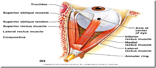
- Superior rectus
- oculomotor nerve
- Inferior rectus
- oculomotor nerve
- Medial rectus
- oculomotor nerve
- Lateral rectus
- abducen nerve
- Superior rectus
- 2 oblique
- Superior oblique
- Inferior oblique
- Levator Palpebrae Superioris (LPS)
- Smooth muscles
- Superior tarsal
- Inferior tarsal
- Orbitalis
- 4 rectus
- Intraocular
- Sphincter pupillae
- Dilator pupillae
- Ciliaris
Bony orbit of the eye
- Superior
- frontal bone
- Inferior
- maxilla
- Medial
- Lacrimal bone
- Lateral
- zygomatic bone
_____________________________________________________________________
Movements of the eyeball
- controlled by the 4 rectus and 2 oblique extraocular muscles
- function
- allow us to follow moving object
- maintain foveal fixation
- binocular vision
- maintain shape of eyeball
- when the movements of 2 eyes are not coordinated
- images of same area of visual fied not focused on same areas of retina
- producing DIPLOPIA
- usually due to weakness/paralysis of extraocular muscles
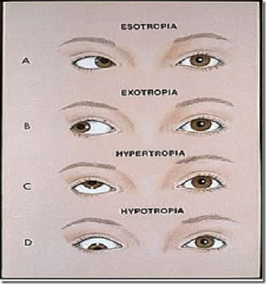
- confenital/acquired, one eye rotated medially/laterally
- producing strabismus/squint
- confenital/acquired, one eye rotated medially/laterally
- images of same area of visual fied not focused on same areas of retina
- function
A – right medial displacement
- muscle paralysed: lateral rectus
B – right lateral displacement
- muscle paralysed: medial rectus
C – superior displacement
D – inferior displacement
_____________________________________________________________________
Monoular movements
- Movement of 1 eye to 1 direction = Duction
- medially: adduction
- laterraly: abduction
- superiorly: supraduction
- inferiorly: infraduction
- Vertical meridian-torsional movement
- nasal rotation, medially along antero-posterior axis
- intortion
- temporal rotation, laterally along antero-posterior axis
- extortion
- nasal rotation, medially along antero-posterior axis
- Agonist
- primary muscle that moves an eye in a given direction
- eg. abduction by lateral rectus
- Synergist
- muscle in the same eye that moves the eye in the same direction as the agonist (support the same movement)
- eg. abduction supported by superior & inferior oblique
- Antagonist
- muscle in the same eye that moves the eye in the opposite direction of the agonist is the antagonist
- eg. medial rectus is an antagonist to abduction by lateral rectus
_____________________________________________________________________
Binocular movements
- Movements are either conjugate (versions) or disconjugate
- movement of 2 eyes in the same direction: version
- Levoversion
- eyes moving in the same direction
- eg. right eye adduction, left eye abducting
- Convergence
- both eyes look towards the nose
- Yoke muscles
- primary muscles in each eye that perform the version
- eg. when looking to the left, left eye perform abduction by lateral rectus muscle. right eye perform adduction by medial rectus muscle. Yoke muscles: lateral and medial rectus muscle.
Visual axis
- Horizontal plane
- Primary plane of action
- Movements are 90 degrees to plane
- Elevation
- Depression
- Vertical plane
- 2ndary plane of action
- Movements are 90 degrees to plane
- Adduction
- Abduction
- Antero-posterior plane
- 3rd plane of action
- Movements are 90 degress to plane
- Intortion
- Extortion
_____________________________________________________________________
Extraocular muscles
Rectus muscles
- Common origin
- All arise from posterior part of orbit
- from a common tendinous ring
- that surrounds optic canal, superior orbital fissure
- All arise from posterior part of orbit
- Insertion into sclera
- inserted into sclera
- 6mm behind sclero-corneal junction
- inserted into sclera
- If not following eyeball plane
- what are the muscles not in allignment?
Levator Palpebrae Superioris (LPS) muscle
- Action: elevates upper eyelid
- Arise from
- Lesser wing of sphenoid
- Partially supplied by
- Somatic nerve (3rd cranial nerve)
- Autonomic (sympathetic – involuntary smooth muscles)
- Superior tarsal
- deep part of LPS
- Inferior tarsal
- connects inferior tarsal plate to fascia around inferior rectus
- both these, innervated by sympathetic nerves = responsible for widely opening the eyelids in response to fear
- Orbitalis
- small smooth muscles bridging inferior orbital fissure
- may compress inferior ophthalmic nerves –making eyeballs prominent
- Superior tarsal
- Aponeurosis divides LPS into 3 layer
- Superficial – skin of upper eyelid
- Middle – tarsal plate of upper eyelid
- Deep – superior conjunctival fornix
Superior Oblique muscle
- Arise from
- body of sphenoid bone
- medial to optic canal
- goes medially, then ends in a tendon
- attached to a pulley: trochlea
- then suddenly runs backward & laterally (change in direction)
- inserted to upper & lateral quadrant of eyeball
- body of sphenoid bone
Inferior oblique muscle
- Arise from
- medial part of floor
- winds round the eyeball laterally & backwards
- inserted to lateral part of sclera
- medial part of floor
_____________________________________________________________________
Movements of the extraocular muscles
| Superior rectus | 1 – elevation 2 – intortion 3 – adduction |
| Inferior rectus | 1 – depression 2 – adduction 3 – extortion |
| Medial rectus | 1- adduction |
| Lateral rectus | 1- abduction |
| Superior oblique | 1 – intortion 2 – depression 3 – abduction |
| Inferior oblique | 1 – extortion 2 – elevation 3 – abduction |
*know the primary, secondary and tertiery movements (refer table)
_____________________________________________________________________
Extraocular muscle disorders
Complete 3rd nerve palsy
- Muscle affected
- medial rectus
- Clinical Presentation
- lateral strabismus
- Medial rectus affected
- diplopia
- eye cannot be moved upwards, medially & downwards
- ptosis
- LPS affected
- *dilated pupil (light reflex sluggish/absent)
- sphincter pupillae affected
- loss of accommodation
- lateral strabismus
- Cause
- diabetes
- atherosclerosis
- tumours
- haemorrhage
4th nerve palsy
- Muscle affected
- Superior oblique
- Diplopia
- difficulty in turning the eyes downwards & laterally while walking down stairs
6th nerve palsy
- Muscle affected
- lateral rectus
- Medial strabismus
- cannot turn eye laterally
- Cause
- facial infection –> cavernous sinus –> infect 6th nerve supplying lateral rectus –> paralysis –> medial strabismus
Extra:
- Sphincter pupillae
- 3rd nerve
- Dilator pupillae
- sympathetic nerve
_____________________________________________________________________
Light reflex
- Useful for gauging brain stem function
- Normally pupils contrict equally
- Direct light reflex
- Each pupil constricts with light shone into that eye
- Consensual pupillary reflex
- Each pupil constricts with light shone into the OTHER eye
 http://library.med.utah.edu/kw/hyperbrain/anim/reflex.html
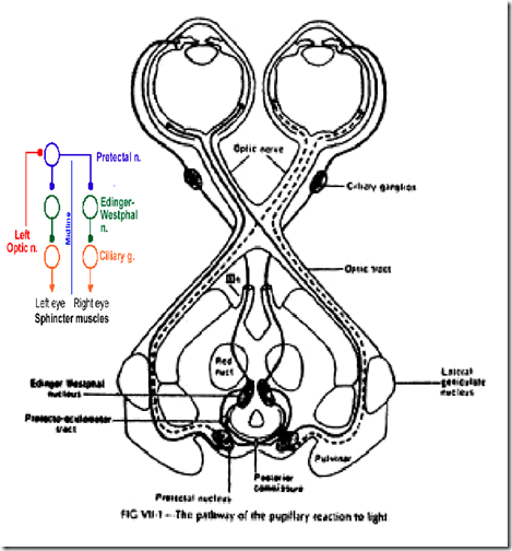
Pupillary light reflex pathway
- Light stimulates retinal photoreceptors
- Afferent fibres transmitted via optic nerve
- Undergo hemi-decussation at optic chiasma
- Exit from optic tract before lateral geniculate body
- lateral geniculate body = vision
- Enters midbrain
- via brachium of superior colliculus
- Synapse at pretectal nucleus
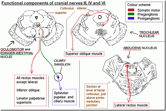
- Fibres go to same-sided & contralateral Edinger –Westphal nuclei
- 3rd nerve
- Efferent fibres travel via 3rd nerve
- Synapse at ciliary ganglion
- Post-ganglionic fibres –> short ciliary nerve
- Innervate left & right sphincter muscles
- constriction of pupil
- Same sided: direct
- Contralateral: consensual
- Pretectal nucleus
- main station for light reflex
- Edinger-Westphal nucleus
- parasympathetic nucleus of oculomotor nerve
- Sphincter muscles
- efferent parasympathetic fibres
- Nerve
- before Edinger-Westphal – Optic nerve (afferent)
- after Edinger-Westphal – Oculomotor nerve (efferent)
Anatomical bases for consensual light reflex
- From pretectal, fibres sent to both Edinger-Westphal nucleus –> pupillary constriction in both eyes
Lack of / abnormal pupillary reflex
- Optic nerve damage
- sensory component
- ipsilateral direct reflex loss
- contralateral direct reflex intact
- Oculomotor nerve damage
- motor component
- damaged side both direct & consensual reflex loss
- opposite side both direct & consensual reflex intact
- Argyll Robertson pupil
- pupil does not react to light, but will react to near accommodation
_____________________________________________________________________
Pathway of sympathetic dilatation of pupil
- Less well-dfined
- Pathway:
- Originates in hypothalamus
- Descends uncrossed to T1
- inferior cervical ganglion
- medial cervical ganglion (stellate)
- superior cervical ganglion
- Exit from spinal cord via perivertebral sympathetic chain
- Synapse in the superior cervical ganglion
- Join ophthalmic division of trigeminal nerve
- via carotid plexus
- Innervate dilator pupillae of the iris
- via the long ciliary nerve
- (short ciliary nerve innervate sphincter pupillae)
- If paralysis
- miosis (constriction of pupil)
- seen in horner’s syndrome
- miosis (constriction of pupil)
_____________________________________________________________________
Accommodation reflex
- Reflex action of the eye in response to focusing on a near object
- Coordinated changes in
- convergence
- medial rectus
- Optic nerve (afferent limb) –> optic tract –> visual cortex –> long association fibre –> frontal eye field –> corticonuclear fibres (pyramidal tract) –> oculomotor nerve (efferent limb) –> contraction of medial rectus of both eyes
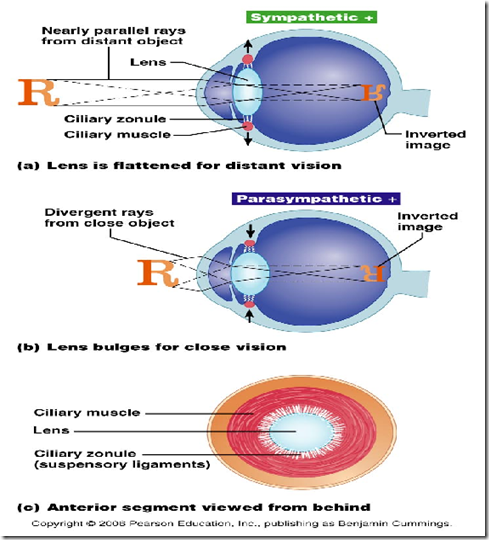
- lens shape
- ciliary muscles
- ciliary muscle contracts, suspensory ligaments relaxed –> making lens more convex – shortening its focal length
- pupil size
- sphincter pupillae
- Edinger-Westphal nucleus –> oculomotor nerve (efferent limb) –> ciliary ganglion –> short ciliary nerve –> ciliary muscle constraction –> sphincter pupillae muscle contraction
- pupil contricts to prevent diverging light rays from hitting the periphery of the retina & producing blurred image
- convergence
- Components
- Afferent: Optic nerve
- Cell station: Multisynaptic
- Efferent: Oculomotor
- Oculomotor nerve supplies these
- smooth muscles
- ciliaris
- sphincter pupillae
- medial rectus
- smooth muscles
_____________________________________________________________________
Other reflexes
Corneal reflex
- Important to detect 7th nerve palsy
- Differential diagnosis: acoustic neuroma
- Components
- Afferent – V1
- Cell station – medial longitudinal fasciculus
- Efferent – Facial nerve (orbicularis)
- Pathway
- light touch of cornea/conjunctiva –> ophthalmic (trigeminal) nerve (Afferent limb) –> sensory nucleus of 5th CN (pons) –> medial longitudinal fasciculus –> motor nucleus of 7th CN –> facial nerve (efferent limb) –> orbicularis oculi (closure of eyelids)
- Which reflex is done to test the 7th CN?
- corneal reflex and conjunctival reflex
- bilateral blinking seen
- Tumour over the 8th CN, can compress the intercostal acoustic meatus (containing 7th CN) –> corneal reflex loss
Visual body reflex
- Optic nerve –> optic tract –> superior colliculus –> tectospinal & tectobulbar tract –> automatic movements of the head, neck & body towards the source of visual stimulus
_____________________________________________________________________
Functional components
Horner’s Syndrome
- Cause
- pancoast tumour compressing the sympathetic nerves at T1 level
- Clinical features
- Ptosis (drooping of eyelid)
- Enophthalmus (reduced prominence of eyeball)
- Anhydrosis
- Miosis (Constriction of pupil)





when afrnt efrnt are all same, why should we check both conjunctival and corneal reflex
Outstanding post but I was wondering if you could write a litte more on
this subject? I’d be very grateful if you could elaborate a little bit further. Cheers!
Attractive section of content. I just stumbled upon your site and in accession capital to assert that I acquire actually enjoyed
account your blog posts. Any way I’ll be subscribing to your
augment and even I achievement you access consistently fast.
If you are going for best contents like I do, simply pay a quick visit this web
site daily because it gives quality contents, thanks